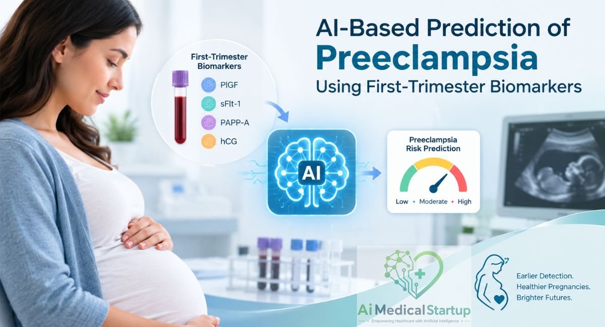

AI-Based Prediction of Preeclampsia Using First-Trimester Biomarkers
Preeclampsia can turn a seemingly normal pregnancy into a serious medical concern, often developing after the first half of pregnancy. The challenge is that early warning signs may be subtle, making timely first-trimester screening especially important for identifying pregnancies that may need closer monitoring. Traditional risk assessment relies on maternal history, blood pressure, ultrasound findings, and selected laboratory measurements, but these factors can interact in complex ways. This is where artificial intelligence in pregnancy is gaining attention.
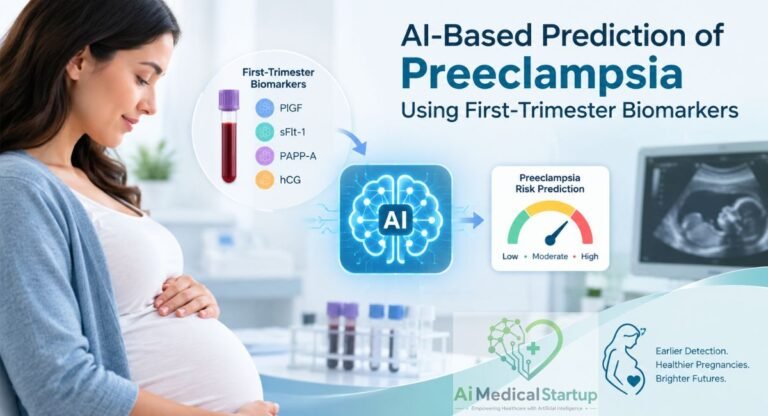
Modern machine learning in healthcare can analyze multiple maternal, biophysical, and biochemical measurements together to uncover patterns that may be difficult to recognize using conventional approaches alone. Biomarkers such as placental growth factor (PlGF), pregnancy-associated plasma protein-A, mean arterial pressure, and uterine artery Doppler measurements can provide valuable information about placental and maternal changes. When combined carefully, these data may support earlier preeclampsia prediction and more personalized prenatal risk assessment.
What Is Preeclampsia and Why Early Prediction Matters
Preeclampsia is a serious pregnancy complication involving new-onset hypertension and other maternal or placental abnormalities. It belongs to the wider group of hypertensive disorders of pregnancy and can affect maternal organs and fetal well-being. Untreated severe disease can contribute to maternal morbidity, preterm birth, fetal growth problems, and serious maternal or perinatal complications.
The tricky part is that clinical disease usually becomes apparent later, while biological changes may develop much earlier. That creates an important window for early preeclampsia prediction and prevention. First-trimester screening aims to identify pregnancies at increased risk, especially for preterm disease. ISSHP recommends screening at 11–14 weeks using clinical factors, blood pressure, uterine artery pulsatility index, and PlGF where available.
What Is Preeclampsia?
Preeclampsia is more than simply having high blood pressure during pregnancy. It can involve maternal organ dysfunction, placental abnormalities, and changes affecting the mother and developing fetus. The condition can become dangerous because its clinical course varies widely. Some pregnancies remain relatively stable, while others develop severe disease requiring early delivery. For readers exploring preeclampsia risk prediction, this distinction matters. Screening estimates the likelihood that disease may develop later. Diagnosis determines whether disease is present now. Therefore, an AI-based prediction of preeclampsia should never be presented as proof that a patient already has the condition.
When Does Preeclampsia Usually Develop?
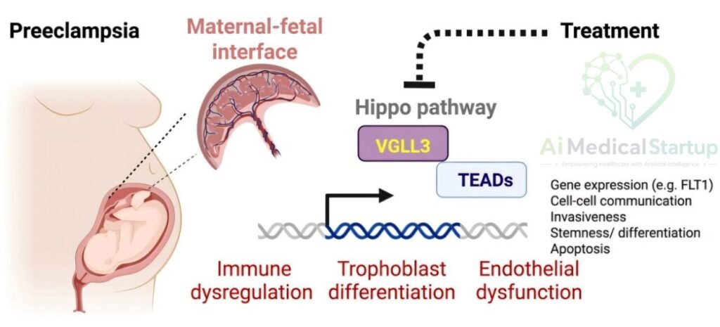
Preeclampsia is generally diagnosed after 20 weeks of pregnancy, although related hypertensive conditions can present differently. Importantly, the biological groundwork may begin much earlier through abnormal placentation, vascular adaptation, inflammation, and endothelial changes. This helps explain why first-trimester preeclampsia prediction has become such an active research area.
Early-onset and late-onset disease may also represent different biological patterns. A prediction system designed for preterm preeclampsia should therefore not automatically be assumed to perform equally well for term disease. That distinction is central to responsible AI-based maternal risk assessment.
Why Is Early Preeclampsia Prediction Difficult?
Preeclampsia does not arise from one simple trigger. Researchers have studied maternal history, blood pressure, placental function, angiogenic proteins, inflammation, metabolic characteristics, and vascular resistance. This creates a complex biological puzzle where individual measurements may provide only part of the picture.
That complexity gives machine learning for preeclampsia prediction an interesting role. Algorithms can examine combinations of variables and identify nonlinear relationships that conventional approaches may not capture easily. Still, a complicated pattern discovered by an algorithm requires clinical validation before it becomes trustworthy.
Why Does First-Trimester Prediction Matter?
The first trimester offers something extremely valuable: time. When clinicians identify increased risk early, they can consider appropriate preventive strategies, monitoring plans, and follow-up according to established guidance. The FMF (Fetal Medicine Archive) describes first-trimester screening using maternal factors, MAP, uterine artery PI, and PlGF to identify women at increased risk for preterm preeclampsia.
This does not mean every woman needs extensive testing. Rather, early pregnancy risk assessment can help distinguish different levels of risk. In the United States, ACOG recommends low-dose aspirin for people at high risk and considers it for certain combinations of moderate-risk factors. Patients should discuss aspirin with their obstetric clinician rather than self-starting treatment.
What Are First-Trimester Biomarkers for Preeclampsia?
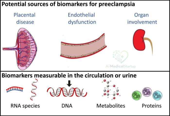
The term first-trimester biomarkers covers measurable biological signals that may provide information about future pregnancy complications. Some come from maternal blood, while others reflect vascular or placental physiology. In practice, researchers often combine pregnancy biomarkers with maternal characteristics and biophysical measurements instead of relying on one laboratory result. This combined approach matters because preeclampsia is biologically heterogeneous. One patient may have strong vascular risk signals, while another may show more pronounced placental or angiogenic abnormalities. By combining maternal biomarkers, clinical information, and biophysical data, prediction models can potentially create a richer picture of pregnancy risk than isolated measurements.
Maternal Characteristics and Clinical Risk Factors
Maternal characteristics form the foundation of many screening systems. Relevant information can include maternal age, previous preeclampsia, chronic hypertension, diabetes, kidney disease, autoimmune conditions, parity, family history, BMI, conception method, and pregnancy type. These maternal risk factors provide context before laboratory measurements enter the equation.
For example, previous preeclampsia can substantially increase future risk, while chronic hypertension, kidney disease, diabetes, and autoimmune disease are also important. ACOG and NICE use structured clinical risk factors when determining who may benefit from preventive strategies.
Blood Pressure and Maternal Hemodynamics
Blood pressure provides another important biological signal. Prediction models often use mean arterial pressure, commonly abbreviated as MAP, because it summarizes arterial pressure across the cardiac cycle. Reliable measurement matters because inconsistent technique can introduce noise into an otherwise sophisticated prediction system.
MAP becomes more informative when combined with other signals. The FMF screening approach, for example, combines maternal factors with MAP, uterine artery PI, and PlGF. This illustrates an important principle: biophysical biomarkers can complement biochemical biomarkers rather than compete with them.
Placental and Angiogenic Biomarkers
Development of the placenta lies at the heart of many preeclampsia models. Placental growth factor, or PlGF, is an angiogenic protein associated with placental vascular development. Other investigated markers include pregnancy-associated plasma protein-A, or PAPP-A, along with soluble fms-like tyrosine kinase-1, commonly called sFlt-1.
These markers should not be treated as interchangeable. Their concentrations change with gestational age and maternal characteristics, while different studies use different combinations. ISSHP recognizes PlGF and several other laboratory measures as investigated early-pregnancy predictors, but also emphasizes that no first- or second-trimester test reliably predicts every case of preeclampsia.
Biochemical and Metabolic Biomarkers
Researchers have investigated many other predictive biomarkers, including inflammatory markers, oxidative-stress signals, metabolic variables, endocrine proteins, and blood-based measurements. Some studies have also examined red blood cell indices and broader laboratory profiles. These variables may capture physiological changes that precede clinically obvious disease.
However, more biomarkers do not automatically create a better model. Adding weak or unstable variables can increase noise and encourage overfitting. Good predictive modeling therefore asks a harder question than “What can we measure?” It asks, “Which measurements consistently improve prediction in independent populations?”
Why Combining Multiple Biomarkers Can Improve Prediction
A single biomarker is like one clue in a detective story. It may be useful, but it rarely explains the entire plot. Combining maternal characteristics, MAP, uterine artery Doppler, PlGF, PAPP-A, and other variables can provide complementary information about placental and maternal physiology.
The FMF reports that combinations of maternal factors, MAP, uterine artery PI, and PlGF can provide substantially stronger detection of preterm disease than maternal factors alone in its screening framework. This is the basic logic behind multimarker screening and increasingly sophisticated algorithmic prediction.
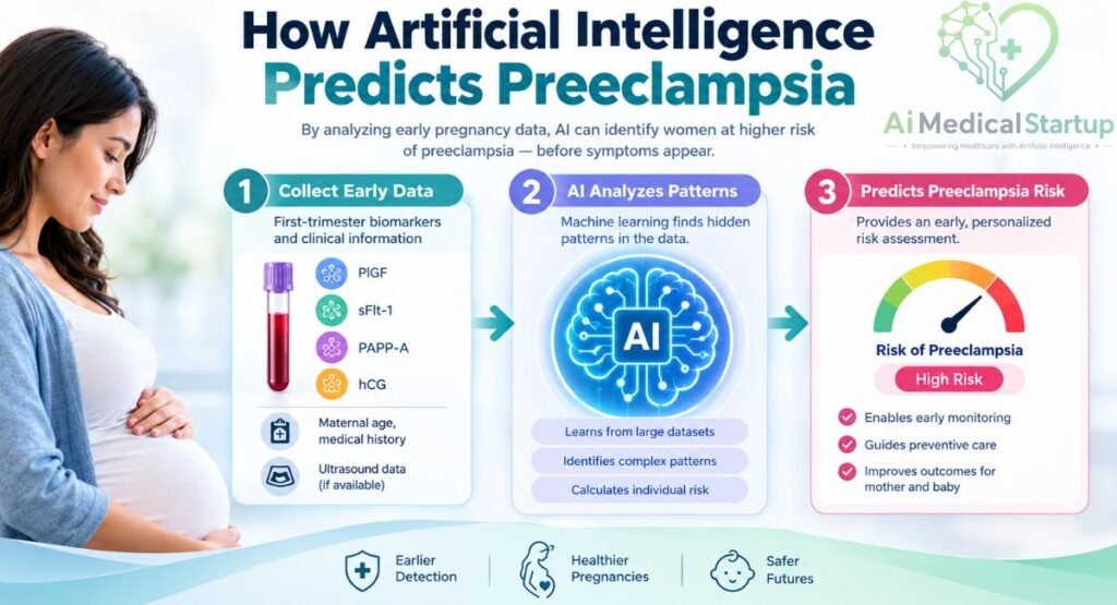
How Artificial Intelligence Predicts Preeclampsia
Artificial intelligence in pregnancy becomes useful when it can transform many measurements into a meaningful risk estimate. Instead of looking at each variable separately, a model learns relationships between inputs and known outcomes. This is one reason AI in obstetrics has attracted interest in early risk prediction, fetal monitoring, imaging, and clinical decision support. Yet AI does not magically understand pregnancy. It learns from the examples researchers provide. If the training data are incomplete, biased, poorly labeled, or too small, the resulting model may learn misleading patterns. In other words, sophisticated software cannot turn poor clinical data into reliable evidence.
Machine Learning vs. Traditional Risk Assessment
Traditional conventional risk assessment often relies on known clinical risk factors and statistical relationships. These methods remain valuable because clinicians understand how they work and because established guidelines can translate risk factors into practical care pathways.
By contrast, machine learning in healthcare can analyze numerous variables and model complex interactions. Machine learning in obstetrics may therefore uncover patterns that are difficult to specify manually. However, a machine-learning model still needs appropriate training, calibration, validation, and clinical interpretation.
How AI Processes First-Trimester Data
An AI system generally starts with structured inputs. These might include maternal demographics, medical history, blood pressure, Doppler measurements, and laboratory markers. The data then pass through preprocessing, feature selection, model training, validation, and final risk estimation.
The process resembles a funnel. Many measurements enter at the top, useful information is refined in the middle, and a smaller output emerges at the end. That output might be a probability or risk category. It is not automatically a diagnosis, prescription, or treatment decision.
Feature Selection and Engineering
Feature engineering converts raw information into variables that a model can use effectively. Researchers may calculate MAP, normalize biomarkers, derive ratios, transform skewed measurements, or express results as gestational-age-adjusted MoM values.
Feature selection then identifies variables that contribute useful predictive information. Methods such as recursive feature elimination, or RFE, can repeatedly remove less useful variables. This can make models faster and sometimes easier to interpret, although feature selection itself must be performed carefully to avoid data leakage.
Managing Missing Data and Class Imbalance
Clinical datasets rarely arrive perfectly clean. Some patients miss laboratory tests, measurements may be recorded differently, and certain outcomes occur less frequently than others. This creates class imbalance, which can cause a model to favor the larger group.
Researchers may use imputation, outlier detection, scaling, and methods such as SMOTE. The synthetic minority oversampling technique creates synthetic examples of the minority class. Importantly, oversampling should occur only within the training data. Otherwise, information can leak into testing and make performance look better than it really is.
Why Data Quality Can Matter More Than Model Complexity
A complicated neural network may look impressive on paper, but it cannot repair unreliable measurements. If gestational age is wrong, biomarker assays vary widely, or outcome labels are inconsistent, the model learns from distorted information.
That is why robust data preprocessing is not housekeeping. It is part of the scientific method. Reliable gestational dating, standardized assays, consistent Doppler measurements, careful outcome definitions, and transparent preprocessing can matter more than adding another algorithm.
Machine Learning Models Used for Early Preeclampsia Prediction
Different algorithms approach the same prediction problem from different angles. Supervised learning models learn from examples where the eventual outcome is already known. In a preeclampsia dataset, the algorithm receives first-trimester measurements alongside information about whether preeclampsia later developed.
No algorithm wins every contest. A logistic regression model may outperform a deep neural network in one dataset, while XGBoost may perform better in another. Model selection should therefore consider sample size, data structure, interpretability, validation, calibration, and clinical usefulness rather than chasing the highest internal score.
Logistic Regression
Logistic regression remains an important baseline because it is relatively interpretable and works well for binary outcomes. It estimates the probability of an event, such as later development of preeclampsia, from selected predictor variables.
Its simplicity is actually an advantage. Researchers can compare more complex models against it and determine whether additional computational complexity provides meaningful improvement. If a complicated model barely beats logistic regression, the simpler approach may be easier to validate and implement.
Random Forest
A random forest, often abbreviated RF, combines many decision trees. Each tree examines different patterns within the data, and their collective predictions create the final output.
This approach can capture nonlinear relationships and interactions without requiring researchers to specify every relationship in advance. However, feature importance from a random forest should not automatically be interpreted as biological causation. The model identifies predictive usefulness, not necessarily disease mechanisms.
Support Vector Machines
A support vector machine, or SVM, classifies observations by identifying useful boundaries between outcome groups. With kernel functions, an SVM can represent complicated relationships that are not easily separated using a straight line.
SVMs can work well with structured datasets, particularly when sample sizes are modest. Their main drawback is interpretability. A clinician may understand the prediction less easily than a straightforward statistical model, especially when several transformations and kernel functions are involved.
Gradient Boosting and XGBoost
Gradient boosting builds an ensemble progressively. Each new tree attempts to improve the errors made by earlier trees. XGBoost is a widely used implementation that can handle structured clinical data and complex relationships.
These models can perform strongly when carefully tuned. Yet aggressive hyperparameter tuning can create an illusion of excellence when datasets are small. The more opportunities researchers have to optimize against the same validation data, the greater the risk of accidentally tailoring the model to that particular sample.
Neural Networks and Deep Learning
A deep neural network, or DNN, contains layers of computational units that can learn complicated nonlinear patterns. Deep learning becomes especially interesting when datasets become larger and include multiple data types.
The attraction is obvious: pregnancy involves interconnected biological processes, and neural networks can model nonlinear interactions. The catch is equally important. DNNs usually need careful training, regularization, sufficient data, and rigorous validation. A high score from a small single-center dataset should never be mistaken for universal clinical accuracy.
Which AI Model Is Best for Preeclampsia Prediction?
There is no universally superior algorithm for preeclampsia prediction. A model that performs beautifully in one hospital may lose accuracy in another because the patient population, laboratory methods, disease prevalence, and clinical practices differ.
Therefore, “best” should mean more than highest accuracy. A clinically useful model should demonstrate discrimination, calibration, reproducibility, model generalizability, interpretability, and ideally external validation across independent populations.
How an AI-Based First-Trimester Prediction System Works
A practical prediction system can be imagined as a clinical pipeline. The patient provides maternal history and routine measurements, while first-trimester testing adds biochemical and biophysical signals. The AI system then processes those variables and produces a risk estimate for later disease.
The important bridge is what happens afterward. A prediction should reach a clinician in a form that can support decision-making. This is where clinical decision support, rather than autonomous treatment, becomes the more realistic goal. The model can flag elevated risk, while the healthcare professional evaluates whether that prediction fits the patient’s circumstances.
| Stage | What happens | Why it matters |
| Patient information | Maternal history and characteristics are collected | Establishes baseline risk |
| Blood pressure | MAP and related measurements are obtained | Captures maternal hemodynamics |
| Biomarkers | PlGF, PAPP-A and other markers may be measured | Adds biological information |
| Doppler | UtA-PI may assess uterine blood flow | Adds placental circulation information |
| Preprocessing | Missing values and measurement differences are addressed | Improves data quality |
| Feature selection | Relevant predictors are retained | Reduces unnecessary noise |
| Model training | Algorithm learns from labeled data | Creates the prediction function |
| Validation | Model is tested on unseen data | Estimates real performance |
| Risk output | Patient receives a probability or category | Supports clinical interpretation |
| Clinical action | Clinician considers monitoring or prevention | Converts prediction into care |
Step 1: Collect Maternal and Pregnancy Data
The first step is deceptively simple. Researchers need accurate maternal information, pregnancy history, gestational age, and clinical measurements. Variables may include maternal age, BMI, parity, previous preeclampsia, chronic hypertension, diabetes, kidney disease, autoimmune conditions, and conception method.
These variables also help contextualize laboratory results. A biomarker value without gestational age or maternal characteristics can be difficult to interpret. This is why first-trimester maternal characteristics often form the first layer of multimarker screening.
Step 2: Measure First-Trimester Biomarkers
The second stage involves laboratory and physiological measurements. Depending on the model, this can include PlGF, PAPP-A, β-hCG, sFlt-1, MAP, and uterine artery Doppler measurements.
Measurement technique matters greatly. The FMF provides detailed protocols for first-trimester uterine artery PI assessment and emphasizes accredited measurement practices for clinical risk assessment.
Step 3: Standardize and Normalize the Data
Raw laboratory values are not always directly comparable. Biomarkers vary with gestational age, maternal characteristics, and laboratory methods. Researchers may therefore convert measurements into multiple of the median, or MoM values, based on appropriate reference distributions.
Normalization helps the algorithm compare signals more consistently. Still, normalization formulas must be transparent. If a model uses one population’s reference medians but is applied to another population, performance can change.
Step 4: Train and Validate the AI Model
During model training, the algorithm learns relationships between predictors and known outcomes. Researchers commonly separate data into training and testing sets and may use fivefold cross-validation during development.
A robust study should preserve a genuinely unseen test set. Otherwise, repeated tuning against the same data can inflate apparent performance. The distinction between internal validation and external validation of AI models is therefore crucial.
Step 5: Generate an Individual Risk Estimate
After training, the system can accept new first-trimester information and produce an estimated probability. The output might say that a pregnancy has relatively low, intermediate, or high predicted risk.
That number needs context. A 10% predicted probability does not mean the patient will definitely develop preeclampsia. It means the model estimates risk under the conditions in which it was developed and validated.
Step 6: Translate the Prediction Into Clinical Action
The final stage should involve a clinician. The result can support AI clinical decision support, but it should not independently prescribe medication or determine delivery timing.
For example, a clinician may combine an elevated model prediction with the patient’s history, examination, guideline recommendations, and available testing. This creates a human-AI partnership rather than an automated obstetrician.
How Accurate Is AI at Predicting Preeclampsia?
Accuracy is often the first number people notice, but it is not the whole story. Preeclampsia prediction accuracy depends on the dataset, outcome definition, prevalence, biomarkers, validation strategy, and prediction threshold. A model can achieve high accuracy while still missing clinically important cases.
For that reason, researchers examine several measures together. Sensitivity, specificity, precision, recall, F1-score, predictive values, and AUC-ROC answer different questions. A trustworthy article should therefore resist the temptation to crown one model based on a single percentage.
Sensitivity and Specificity
Sensitivity measures how well a model identifies people who eventually develop the target condition. Specificity measures how well it identifies people who do not. In screening, both matter because false negatives and false positives have different consequences.
A model with high sensitivity may catch more high-risk pregnancies but could also generate more false alarms. Conversely, a highly specific model may reduce unnecessary alerts while missing some cases. The best balance depends on clinical purpose and acceptable risk.
Accuracy, Precision, and F1 Score
Prediction accuracy describes the proportion of correct classifications, but it can become misleading when one outcome is much more common. Precision asks how many predicted positive cases were actually positive, while recall is another name for sensitivity.
The F1-score combines precision and recall into one measure. It can be useful when researchers want a balance between identifying true cases and limiting incorrect positive predictions.
Area Under the ROC Curve (AUROC)
The area under the ROC curve, or AUC-ROC, measures discrimination across different classification thresholds. An AUC closer to 1 generally indicates stronger ability to distinguish higher-risk from lower-risk cases within the evaluated dataset.
However, AUC does not tell you whether the probabilities are well calibrated. Nor does it prove that using the model improves maternal outcomes. ROC curve analysis is useful, but it is only one piece of the evidence puzzle.
Positive and Negative Predictive Values
The positive predictive value tells you how many people classified as high risk actually develop the condition. The negative predictive value tells you how many classified as low risk remain disease-free.
Unlike sensitivity and specificity, predictive values depend strongly on disease prevalence. A model can therefore have different PPV and NPV when transported from a specialist research cohort to a general pregnancy population.
Why Model Performance Can Differ Between Studies
Two studies can use the same algorithm and obtain very different results. One may recruit a high-risk hospital population, while another uses a broad community cohort. Their laboratory methods, biomarker thresholds, outcome definitions, and missing-data strategies may also differ.
This is why model performance must always be interpreted alongside study design. A 95% accuracy result from a small retrospective cohort does not mean the same model will achieve 95% accuracy in every hospital, country, or demographic group.
Why a High Accuracy Score Does Not Guarantee Clinical Readiness
Clinical readiness requires more than discrimination. Researchers should consider calibration, external validation, prospective testing, decision thresholds, clinical workflow, patient acceptability, and potential harms.
A model can correctly rank high-risk patients yet provide poorly calibrated probabilities. Another can perform well statistically but prove difficult to integrate into routine care. Clinical utility of AI is therefore broader than mathematical performance.
Which Biomarkers Contribute Most to AI Preeclampsia Prediction?
Some variables repeatedly appear in early-pregnancy prediction research because they capture important aspects of maternal or placental physiology. PlGF, PAPP-A, MAP, and uterine artery PI are particularly relevant in established multimarker screening frameworks.
Still, “important” does not mean “diagnostic.” A variable can improve prediction because it contains useful statistical information without directly causing disease. This distinction becomes especially important when interpreting biomarker feature importance from machine-learning models.
Placental Growth Factor and Angiogenic Markers
Placental growth factor preeclampsia research has attracted considerable attention because PlGF reflects placental angiogenic activity. The FMF incorporates PlGF into its first-trimester screening framework alongside maternal factors, MAP, and uterine artery PI.
The related PlGF preeclampsia prediction concept should still be interpreted carefully. PlGF is one component of a risk model, not a stand-alone diagnostic test for future disease. Other angiogenic markers, including sFlt-1, are also studied, particularly later in pregnancy.
Pregnancy-Associated Plasma Protein-A
PAPP-A is measured during first-trimester screening and has been investigated as one of several preeclampsia biomarkers. Researchers have explored whether altered PAPP-A levels provide information about placental development and later pregnancy complications.
The important point is context. The FMF’s published screening tables show that adding PAPP-A to combinations containing PlGF may not provide substantial additional detection for preterm disease in its framework. Therefore, PAPP-A should not automatically be presented as superior simply because it is widely available.
Uterine Artery Doppler Measurements
Uterine artery Doppler preeclampsia assessment adds a physiological dimension to blood and demographic data. The uterine artery pulsatility index, or UtA-PI, provides information about resistance to blood flow in the uterine arteries.
The measurement requires technical consistency. FMF guidance specifies first-trimester timing and a standardized Doppler technique, including identification of the uterine arteries and calculation of the mean PI. This matters because poor measurement quality can undermine even the best algorithm.
Blood Pressure and Maternal Factors
MAP preeclampsia prediction works because maternal hemodynamics provide information that biomarkers alone cannot capture. Likewise, BMI and preeclampsia risk, maternal age, previous pregnancy history, chronic disease, and other variables can alter baseline probability.
This is why AI systems often perform better when they combine biological signals with clinical context. A low PlGF value means something different depending on gestational age, maternal characteristics, and the rest of the clinical profile.
Combining Biomarkers With Clinical Data
The strongest conceptual model is multimodal. Instead of asking which single biomarker predicts preeclampsia, researchers ask how different signals work together.
The FMF framework is a useful example because it combines maternal factors, MAP, UtA-PI, and PlGF. Its published data show substantially higher detection of preterm disease with combined markers than maternal factors alone.
Making AI Preeclampsia Models Explainable
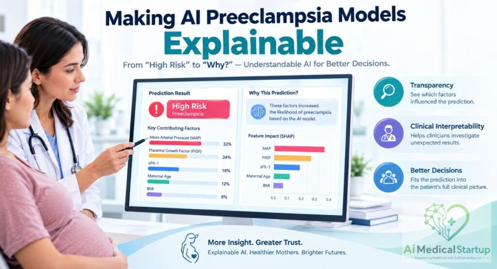
A prediction that simply says “high risk” can leave clinicians asking a natural question: why? Explainable AI in obstetrics attempts to answer that question by showing which variables contributed most strongly to an individual or overall prediction. This matters because pregnancy decisions involve real people, not abstract rows in a spreadsheet. AI transparency, clinical interpretability, and understandable explanations can help clinicians investigate unexpected results and decide whether a prediction fits the patient’s broader clinical picture.
What Is Explainable AI in Healthcare?
Explainable artificial intelligence in healthcare refers to methods that help humans understand how a model reaches its predictions. Some models are inherently easier to interpret, while others require additional explanation techniques.
For clinicians, the goal is not to understand every mathematical operation. Instead, they need meaningful information about the factors influencing the prediction and the confidence or limitations surrounding it.
How SHAP Can Identify Important Biomarkers
SHAP analysis, short for SHapley Additive exPlanations, is one method researchers use to interpret machine-learning models. SHAP values can estimate how individual features contribute to a particular prediction.
For example, a model may identify PlGF, MAP, UtA-PI, BMI, or maternal age as influential variables. However, feature importance does not prove causality. The model is showing predictive contribution, not demonstrating that changing the variable will prevent preeclampsia.
Why Clinicians Need Interpretable AI Predictions
Imagine receiving a laboratory result that simply says “high risk” with no explanation. You would probably want to know what produced that result. Clinicians are no different.
Interpretability can support error checking, communication, auditing, and clinical reasoning. It may also help reveal unexpected patterns, such as an algorithm relying heavily on a variable that reflects a hospital’s documentation habits rather than true disease biology.
From Black-Box Prediction to Clinical Decision Support
The most useful future role for AI may be as an intelligent assistant. A model could process complex information, flag elevated risk, and show the major contributing factors.
The clinician would then combine that output with examination findings, guidelines, patient preferences, and additional investigations. This model respects the difference between algorithmic prediction and medical judgment.
Why Feature Importance Does Not Prove Causation
A feature can be highly predictive without causing the disease. For example, a variable may act as a proxy for another physiological or demographic characteristic.
Therefore, researchers should avoid statements such as “AI discovered that biomarker X causes preeclampsia.” Prediction and causation are different scientific questions. AI trust depends partly on maintaining that distinction.
Potential Benefits of AI-Based Early Preeclampsia Prediction
The strongest argument for AI is not that computers are clever. It is that early, structured risk information could help clinicians organize care more intelligently.
If validated properly, AI-assisted screening could combine information that is otherwise scattered across medical history, laboratory systems, ultrasound reports, and vital-sign measurements. That could support personalized prenatal care while reducing the cognitive burden of manually interpreting numerous variables.
Earlier Identification of High-Risk Pregnancies
Earlier identification creates more time for appropriate management. The FMF describes first-trimester screening specifically as a way to identify pregnancies at increased risk of preterm preeclampsia.
However, identification should not be confused with certainty. The output is a risk estimate. A high-risk prediction means closer consideration may be appropriate, not that preeclampsia is inevitable.
More Personalized Prenatal Monitoring
Not every pregnancy carries the same baseline risk. Personalized pregnancy management could use validated risk estimates to help determine how closely a pregnancy should be followed.
That does not mean replacing routine prenatal care. Instead, risk stratification may help clinicians decide when additional surveillance, laboratory testing, blood-pressure monitoring, or specialist review deserves greater attention.
Supporting Preventive Interventions
Early screening can potentially support evidence-based preventive interventions. Low-dose aspirin is one example, but the decision should follow appropriate clinical guidance rather than an AI output alone.
ACOG recommends low-dose aspirin for pregnant individuals with specified high-risk factors and for some combinations of moderate-risk factors. It also advises patients to discuss aspirin with their obstetric clinician rather than taking it independently.
Improving Maternal and Fetal Outcomes
The ultimate goal is better maternal and fetal outcomes, not a prettier ROC curve. Earlier risk identification could theoretically support timely monitoring and intervention, particularly when disease threatens maternal health or requires preterm delivery.
Still, outcome improvement must be demonstrated. A model with impressive predictive statistics does not automatically reduce maternal mortality, perinatal morbidity, or other adverse outcomes. Clinical trials and real-world evaluations are needed to establish that link.
Reducing Unnecessary Monitoring for Lower-Risk Patients
Risk stratification can potentially work in both directions. Identifying higher-risk pregnancies may focus resources where they are most needed, while reliable low-risk estimates could potentially reduce unnecessary investigations.
That benefit requires excellent calibration. If a supposedly low-risk model misses important cases, the result could be harmful. Therefore, automated risk assessment should be implemented cautiously and monitored continuously.
Why AI Could Matter in Resource-Limited Healthcare Systems
A validated digital model could eventually help standardize risk assessment where specialist resources are scarce. AI implementation in resource-limited settings could potentially support clinicians by organizing complex information and highlighting patients who may require additional review.
However, technology is not automatically affordable or accessible. Reliable internet, laboratory testing, ultrasound expertise, data infrastructure, maintenance, cybersecurity, and clinician training all influence whether scalable AI systems can actually work.
Limitations and Challenges of AI Preeclampsia Prediction
AI prediction sounds exciting, but medicine has a habit of punishing overconfidence. A model can achieve excellent internal performance and still fail when exposed to new patients.
The biggest challenge is model generalizability. Preeclampsia differs across populations, healthcare systems, laboratories, and clinical settings. A model trained in one hospital may encounter very different patients elsewhere. This makes external testing essential before widespread implementation.
Small or Biased Datasets
Small datasets can make AI models appear more accurate than they truly are. When researchers test many algorithms and parameters against limited data, the model may learn quirks of the sample rather than general disease patterns.
Selection bias can create another problem. A specialist hospital may see more severe cases than a general population. A model trained there could perform poorly in routine community prenatal care.
Differences Between Hospitals and Populations
Population differences can strongly influence prediction. Maternal age, BMI, disease prevalence, social conditions, healthcare access, laboratory methods, and clinical practice all vary.
The FMF itself notes that screening performance and screen-positive rates can differ between populations. This is one reason multi-center validation matters before assuming that one prediction model fits everyone.
Biomarker Availability and Cost
Some biomarkers and Doppler measurements require equipment, trained staff, laboratory infrastructure, and quality control. That can create barriers to adoption.
A model that depends on expensive testing may be difficult to deploy in low-resource environments. Conversely, a simpler model using routinely collected information might be easier to scale, even if its predictive performance is somewhat lower.
Overfitting and Model Generalization
Overfitting happens when a model becomes too tailored to its training data. It may memorize patterns that look useful internally but disappear in new patients.
Strong cross-validation, an untouched test set, transparent preprocessing, and external validation of AI models can reduce this problem. Prospective studies are even more valuable because they test the model under real clinical conditions.
Data Privacy and Ethical Concerns
Pregnancy data can contain highly sensitive medical information. AI systems may combine laboratory results, medical histories, imaging, demographic characteristics, and electronic health records.
Responsible AI implementation in healthcare therefore requires strong privacy protections, secure data handling, appropriate consent, bias monitoring, and clear accountability. A technically excellent model can still be unacceptable if it mishandles patient information.
The Need for External and Prospective Validation
External validation asks whether the model works in an independent population. Prospective validation goes further by testing predictions on future patients under predefined conditions.
These steps help answer the question that matters most: does the model still work when nobody has already seen the answers? Without that evidence, promising AI model performance remains preliminary.
What Does the Future Hold for AI-Based Preeclampsia Prediction?
The next generation of artificial intelligence in maternal health will probably become more multimodal. Instead of relying on one blood test or one clinical visit, future systems may combine laboratory results, imaging, electronic records, wearable measurements, and longitudinal observations.
This could move the field toward precision obstetrics. Rather than asking whether a patient is simply “high risk” or “low risk,” AI could potentially estimate changing risk over time and identify different biological patterns. That vision remains promising, but it requires stronger datasets and careful prospective research.
Multimodal AI Using Biomarkers, Imaging, and Clinical Records
Multimodal AI can combine different types of information. A future preeclampsia model might integrate maternal characteristics, biomarkers, Doppler measurements, ultrasound features, blood-pressure readings, and clinical records.
This approach may reveal relationships that remain invisible when each data source is analyzed separately. However, multimodal systems also create new challenges. Missing data, incompatible formats, privacy concerns, and complex validation become harder when more information enters the model.
Integration With Electronic Health Records
EHR integration could allow prediction models to access information already collected during prenatal care. Instead of asking clinicians to enter the same information into another system, an integrated model could retrieve appropriate variables automatically.
That convenience could improve adoption. Yet automatic data extraction introduces its own risks. Incorrect coding, outdated medication lists, missing histories, and inconsistent documentation can silently affect an algorithm’s output.
Real-Time Pregnancy Risk Monitoring
Future systems may move beyond one-time screening toward real-time AI clinical prediction. Blood-pressure measurements from home devices, laboratory results, symptoms, and other longitudinal signals could potentially update risk over time.
This approach resembles a weather forecast more than a one-time diagnosis. The forecast changes as new information arrives. Similarly, pregnancy risk may evolve, meaning an AI system could potentially update estimates as the pregnancy progresses.
Personalized Risk Prediction
Personalized prediction is one of the most attractive ideas in precision pregnancy care. Two patients can have similar blood pressure but very different histories, biomarker patterns, and baseline risks.
A sufficiently validated model could account for these differences. Yet personalization should never become an excuse for opaque decision-making. Clinicians and patients still need understandable information about why a prediction changed.
AI as a Clinical Decision-Support Tool
The most realistic near-term role for AI may be AI clinical decision support rather than autonomous obstetric care. A system could summarize risk, identify relevant variables, flag unusual patterns, and remind clinicians when additional assessment may be appropriate.
This approach keeps humans in the loop. It also creates a safer division of responsibility: the algorithm processes complexity, while clinicians interpret the result within the patient’s real-world context.
Combining AI With Genomics and Multi-Omics
Future research may add genomic data, transcriptomic data, proteomic information, and metabolic profiles. These layers could reveal molecular signatures associated with placental dysfunction and different preeclampsia phenotype.
However, more data can also mean more noise. The challenge will be identifying information that genuinely improves prediction rather than simply making the model larger.
From Single-Timepoint Prediction to Longitudinal AI
Longitudinal biomarkers could allow researchers to examine how biological signals change during pregnancy. Instead of asking what one measurement means at 12 weeks, a model could potentially study trajectories across several time points.
That could be particularly valuable because preeclampsia is dynamic. A changing biomarker pattern may carry information that a single measurement cannot capture. This remains an important research direction rather than established routine care.
AI vs. Traditional Preeclampsia Risk Prediction
Traditional screening remains the foundation of pregnancy care. Clinical history, blood pressure, examination, laboratory testing, ultrasound, and established guidelines already provide valuable information. AI should therefore be viewed as a potential extension of conventional preeclampsia prediction, not an automatic replacement.
The Fetal Medicine Foundation algorithm is an important example of structured multimarker screening. It combines maternal factors with measurements such as MAP, UtA-PI, and PlGF to estimate risk. The comparison below shows where AI may add value and where traditional methods remain essential.
| Feature | Traditional Risk Assessment | AI-Based Prediction |
| Maternal history | Strong role | Can be integrated |
| Blood pressure | Routinely used | Can be integrated |
| Biomarkers | Used in selected screening systems | Can combine many variables |
| Doppler measurements | Established role | Can be incorporated |
| Complex interactions | More limited | Strong computational potential |
| Large datasets | Manual analysis is difficult | Well suited to computation |
| Personalized risk | Possible | Potentially more granular |
| Interpretability | Usually straightforward | Depends on model |
| External validation | Important | Essential |
| Clinical decision support | Established | Emerging |
| Autonomous treatment | Not appropriate | Not appropriate |
Does AI Replace Traditional Preeclampsia Screening?
No. At least not based on current evidence. Existing screening approaches have clinical foundations, standardized protocols, and guideline frameworks. AI models must prove that they improve meaningful outcomes before they can reasonably displace established methods.
The ISSHP explicitly notes that no first- or second-trimester test can reliably predict every case of preeclampsia. That caution is important because even excellent screening systems have limitations.
AI Screening vs. Clinical Diagnosis
Screening asks, “Who is more likely to develop this condition?” Diagnosis asks, “Does this patient have the condition now?” Risk stratification sits between those concepts and organizes patients according to predicted likelihood.
That distinction should remain clear throughout any discussion of AI-driven preeclampsia prediction. A machine-learning probability cannot independently diagnose preeclampsia, decide delivery timing, or replace clinical assessment.
Frequently Asked Questions About AI and Preeclampsia
Can AI predict preeclampsia in the first trimester?
Yes, AI models can be developed to estimate first-trimester risk using maternal characteristics, blood pressure, Doppler findings, and biochemical measurements. However, early prediction of preeclampsia is probabilistic, not certain. Current evidence also shows that no early test reliably predicts every case.
Which biomarkers are associated with preeclampsia risk?
Several biomarkers for preeclampsia prediction have been investigated, including PlGF, PAPP-A, sFlt-1, β-hCG, inflammatory markers, and other laboratory signals. Their usefulness depends on gestational age, population, assay methods, and the prediction model in which they are used.
How accurate are AI models for preeclampsia prediction?
Reported performance varies widely between studies. Sensitivity and specificity of AI models, AUC, calibration, predictive values, and external validation all matter. A high internal accuracy score should not be interpreted as proof that a model will perform equally well in another hospital or population.
Can machine learning replace traditional preeclampsia screening?
Current evidence does not justify assuming that machine learning can replace established screening. Instead, machine learning in preeclampsia may complement clinical risk assessment by combining multiple variables and identifying complex patterns. Any replacement would require strong comparative and prospective evidence.
Is AI-based preeclampsia prediction available in clinical practice?
Some structured risk calculators and multimarker screening approaches already exist, but that does not mean every research AI model is clinically validated. The FMF provides a preeclampsia risk-assessment framework based on maternal factors and biomarker measurements, with requirements around appropriate measurement and practitioner competence.
Can AI predict early-onset and late-onset preeclampsia separately?
Potentially, yes. Researchers can train models around different disease endpoints, such as early-onset or preterm preeclampsia. However, performance should be evaluated separately because these phenotype may have different biological characteristics and prevalence.
Can AI predict preeclampsia before symptoms appear?
AI can estimate future risk before clinical disease becomes apparent when appropriate first-trimester variables are available. The important phrase is “estimate future risk.” Early detection of preeclampsia is not the same as diagnosing disease before symptoms.
Can biomarkers alone predict preeclampsia?
Biomarkers can contribute valuable information, but no single first-trimester biomarker reliably predicts every case. Multimarker approaches often combine biochemical, biophysical, and maternal information to improve risk estimation.
What is the role of PlGF in preeclampsia prediction?
PlGF is an important angiogenic marker used in established screening frameworks. The FMF combines PlGF with maternal factors, MAP, and UtA-PI for first-trimester risk assessment of preterm disease.
What is the role of PAPP-A in first-trimester screening?
PAPP-A is a pregnancy-associated protein measured in first-trimester screening. It has been investigated as a predictor of preeclampsia and other pregnancy outcomes. Its incremental value depends on the screening model and biomarkers already included.
Conclusion: Can AI Make Preeclampsia Prediction Earlier and More Personalized?
The promise of artificial intelligence-based prediction of preeclampsia using first-trimester biomarkers lies in integration. AI can potentially bring together maternal characteristics, MAP, uterine artery Doppler, PlGF, PAPP-A, and other clinical measurements into one structured risk estimate. That could make early pregnancy risk assessment more individualized.
However, the smartest algorithm is not automatically the safest one. Reliable AI-based prediction of preeclampsia requires representative data, rigorous preprocessing, transparent methods, external validation, prospective evaluation, and meaningful clinical utility. The future is therefore unlikely to be “AI versus doctors.” It is more likely to be clinicians using well-validated AI as another tool for earlier, clearer, and more personalized pregnancy care.


Dr. Kanza Sarfraz, M.B.B.S., is a medical doctor and graduate of Allama Iqbal Medical College, Lahore. She brings nearly seven years of clinical experience across tertiary-care hospitals, medical headquarters, and healthcare facilities in both the public and private sectors. Her clinical experience provides a practical perspective on healthcare delivery, emerging medical technologies, and the evolving role of artificial intelligence in medicine.