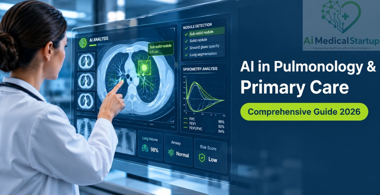
AI in Pulmonology

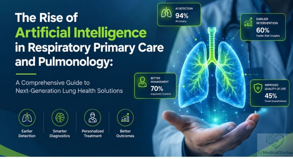
The Rise of AI in Respiratory Care
A Comprehensive Guide to Next-Gen Pulmonology & Primary Care Solutions
Artificial intelligence is reshaping modern healthcare, and Respiratory medicine is becoming one of its most promising fields of transformation. Across primary care and pulmonology, intelligent technologies are helping clinicians interpret complex patient data, identify subtle disease patterns, and make faster clinical decisions. From advanced Diagnostic imaging to automated lung function analysis, AI can support earlier recognition of conditions that might otherwise remain unnoticed until symptoms become severe. This shift is particularly important as healthcare systems face rising respiratory disease burdens, growing patient volumes, and persistent workforce shortages.
AI-powered tools can help bridge gaps between frontline primary care and specialized pulmonary expertise while supporting more consistent evaluation across different clinical settings. At the same time, Pulmonary care is moving toward more proactive and personalized management, with digital systems capable of monitoring patients beyond traditional appointments. This comprehensive guide explores how AI is transforming respiratory diagnosis, treatment, monitoring, research, and the future of next-generation lung health solutions.
The Evolving Landscape of Respiratory Medicine
The practice of modern Respiratory medicine is moving rapidly away from episodic reactive care toward continuous physiological oversight. Primary health hubs now utilize cloud-connected diagnostic devices, automated clinical notes tools, and deep imaging models to track subtle disease progression in real time. This structural transformation relieves pressure on overcrowded emergency rooms by identifying physiological declines early and keeping patients out of the hospital.
Evolution of AI Techniques in Pulmonology
Classical Machine Learning: The Era of Feature Engineering
Early medical software relied on structured statistical models and manual data extraction techniques to analyze patient parameters. Classical Machine Learning (ML) approaches, such as Support Vector Machines (SVM) and Random Forests (RF) / Ensemble Learning, successfully classified basic pulmonary function metrics by processing curated demographic and physiological inputs. These early implementations established foundational concepts for digital risk scoring in clinical settings, proving that mathematical algorithms could reliably flag abnormal lung dynamics.
Deep Learning: Mastering Unstructured Clinical Data
The arrival of Deep Learning (DL) architectures revolutionized medical analysis by extracting intricate patterns directly from raw physiological measurements and complex pixel arrays. Modern diagnostic systems utilize Convolutional Neural Networks (CNNs / 2D/3D CNN) alongside spatial Transformers (Vision Transformer [ViT], Swin Transformer) to automatically map spatial relationships inside complex thoracic scans without requiring hand-coded rules. Neural networks using backbone architectures like U-Net / ResNet / DenseNet segment delicate lung zones, highlight structural boundaries, and detect tissue density changes in seconds.
Generative AI, LLMs, and Foundation Models: The Current Frontier
Generative architectures and specialized Med-LLMs (e.g., Med-PaLM) represent the cutting edge of clinical automation, converting complex raw patient files into concise clinical overviews. Modern Multimodal foundation models and Generalist Medical AI (GMAI) engines process structured diagnostic numbers, raw imaging scans, and text entries simultaneously. These systems apply Natural Language Processing (NLP) and Cross-modal reasoning to analyze longitudinal patient charts and automatically generate comprehensive Report summarization outputs for multidisciplinary medical teams.
Key Drivers: Diagnostic Delays, Workforce Shortages & Data Overload
Systemic delays in primary care clinics across North America and Europe often result in late-stage diagnoses for critical conditions like lung cancer and idiopathic pulmonary fibrosis. General practitioners are overwhelmed by massive administrative workloads, reviewing hundreds of complex medical records and diagnostic images daily. Algorithmic software systems relieve this operational pressure by automatically triaging routine cases and alerting care teams immediately to high-risk physiological abnormalities.
AI in Respiratory Diagnostic Imaging
Thoracic radiology generates massive amounts of complex visual data that stretch clinical bandwidth past sustainable limits. Deploying automated Diagnostic imaging algorithms directly into primary screening pipelines speeds up patient evaluation by instantly analyzing chest radiographs and high-resolution computed tomography scans. Computer vision software acts as an tireless assistant for clinicians, flagging microscopic structural changes during routine checkups.
Computer vision models minimize subjective interpretation variances between different healthcare facilities and rural clinics. Algorithms analyze pixel density variations to identify subtle tissue changes that might escape notice during high-volume manual reviews. Primary care practices equipped with visual screening tools can deliver expert-level imaging assessments right in the local community.
Chest Radiography and Computed Tomography (CT)
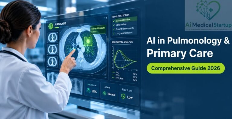
Early Lung Cancer Detection and Nodule Characterization Identifying sub-solid Pulmonary nodules on low-dose computed tomography scans is critical for successful long-term outcomes in Thoracic oncology. Advanced Deep Learning (DL) models process three-dimensional scans to measure exact volumetric growth, evaluate border tissue characteristics, and calculate malignancy risks automatically.
Diagnostic workflow:
Multi-slice volumetric imaging input from low-dose chest CT scans.
Automated isolation of parenchymal tissue and sub-solid nodules.
Temporal comparison of lesion growth rates across prior scans.
Calculated probability score to assist multidisciplinary care teams.
These automated risk scores help clinicians follow standardized medical guidelines, ensuring patients with suspicious growths get referred for biopsies quickly without subjecting low-risk individuals to unnecessary invasive testing.
Interstitial Lung Disease (ILD) and COPD Phenotyping
Quantifying structural tissue degradation in Interstitial lung diseases (ILDs) requires detailed density mapping across multiple anatomical zones. Advanced image algorithms segment ground-glass opacities, honeycombing structures, and emphysematous tissue loss in seconds.
Honeycombing Index
Quantifies permanent fibrotic tissue damage throughout lower lung fields.
Emphysema Rating
Calculates low-attenuation areas below minus nine hundred fifty Hounsfield units to assess destructive Chronic obstructive pulmonary disease (COPD) changes.
Airway Diameter Tracking
Measures lumen narrowing and wall thickening across distal airways.
Acute, Infectious, and Dynamic Diagnostics
Automated Detection of Pneumonia, Tuberculosis, and COVID-19
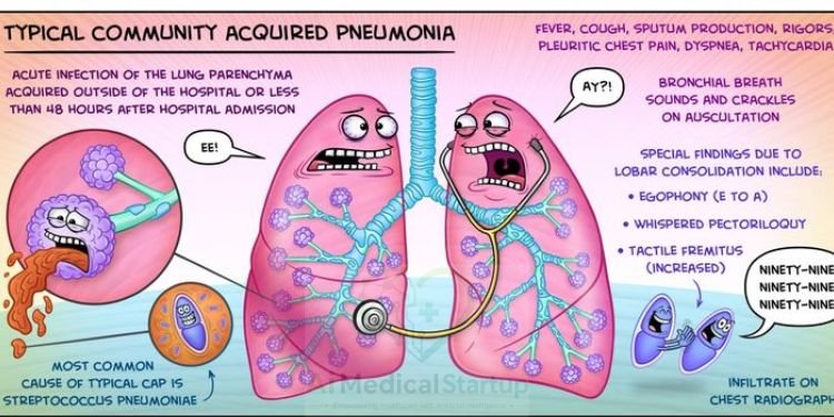
Rapid evaluation tools analyze standard chest radiographs to identify consolidation patterns caused by acute bacterial or viral pathogens. High-speed algorithms for Tuberculosis (TB) screening and COVID-19 / pneumonia detection accelerate triage in community health centers, isolating infectious cases quickly and stopping community spread.
Point-of-Care Ultrasound (POCUS) Integration
Handheld ultrasound devices enhanced with real-time computer vision guide primary care doctors through bedside thoracic evaluations. “Integrating artificial intelligence into point-of-care ultrasound empowers primary care teams to perform high-precision thoracic examinations at the bedside, delivering accurate fluid estimates without waiting for formal radiology scans.”
Automated B-line counting and pleural fluid volume estimates allow instant diagnosis of acute lung congestion or fluid build-up during routine outpatient visits.
Pulmonary Angiography and Vascular Imaging (Pulmonary Embolism Detection)
Computer vision frameworks evaluate computed tomography angiography scans to identify dangerous filling defects within pulmonary arteries. Automated detection algorithms notify emergency response teams instantly, drastically cutting time-to-treatment for high-risk vascular blockages and reducing sudden mortality risks.
AI in Pulmonary Function Testing (PFTs) and Physiological Signals
Evaluating airflow limitations through lung function testing requires patient cooperation and precise interpretation. Intelligent software platforms evaluate test effort quality in real time, comparing raw effort loops against global respiratory database standards. These automated tools bring specialized clinical expertise directly to local primary care facilities.
Integrating automated signal analysis turns complex breath-by-breath recordings into clear diagnostic insights. Software algorithms eliminate common interpretation errors by identifying poor patient effort, mask leak artifacts, and subtle early airway obstructions. This continuous oversight ensures dependable diagnostic readings across every clinic.
Automated Interpretation of Spirometry and Plethysmography
Diagnostic engines process flow-volume curves to distinguish restrictive lung conditions from obstructive disorders like Chronic obstructive pulmonary disease (COPD).
| Testing Metric | Normal Physiological Range | Obstructive Airway Pattern | Restrictive Tissue Pattern |
|---|---|---|---|
| FEV1 / FVC Ratio | Greater than or equal to 0.70 | Less than 0.70 | Normal or elevated value |
| Total Lung Capacity | 80 percent to 120 percent predicted | Normal or hyperinflated volume | Less than 80 percent predicted |
| Diffusing Capacity (DLCO) | 80 percent to 100 percent predicted | Variable based on emphysema | Reduced in parenchymal disease |
Digital Stethoscopes and Acoustic AI for Cough/Wheeze Analysis
Smart digital stethoscopes record body soundscapes to classify abnormal breath sounds, such as fine fibrotic crackles and high-pitched wheezes. Acoustic models filter out environmental noise to isolate subtle sound frequencies, helping general practitioners confirm early airway constriction or fluid buildup before severe symptoms appear.
Cardiopulmonary Exercise Testing (CPET) Data Analytics
Algorithms evaluate breath-by-breath gas exchange data to distinguish heart-related exercise limits from lung-related ventilatory restrictions.
Diagnostic pathway:
Cardiac Exercise Restriction
- Low O₂ Pulse
- Inadequate Heart Rate Response
Ventilatory Exercise Restriction
- Elevated VE/VCO₂ Slope
- Low Breathing Reserve (BR)
Automated data analytics identify the precise physiological bottleneck limiting a patient's physical endurance during structured exertion tests.AI in Treatment Planning and Clinical Decision Support (CDSS)
Choosing the right treatments for chronic lung conditions requires matching current medical guidelines with individual patient traits. Modern Clinical Decision Support Systems (CDSS) compare local patient profiles against massive clinical treatment databases to recommend tailored therapeutic strategies. These decision platforms help family doctors manage complex respiratory treatments safely.
By cross-referencing patient charts with updated medical research, decision engines help prevent adverse drug interactions and highlight emerging treatment resistance. Intelligent software keeps community practice patterns aligned with global treatment protocols.
Precision Oncology and Interventional Decision Support
Target Volume Delineation in Radiotherapy: Deep learning models outline tumor boundaries and surrounding healthy organs on planning CT scans within minutes.
Treatment Speed
Reduces manual contouring times from hours to minutes for oncology specialists.
Target Accuracy
Eliminates outline variations between different radiation oncologists.
Healthy Tissue Protection
Spares functional lung tissue from unnecessary radiation exposure.
Robotic Bronchoscopy and AI-Guided Biopsies
Intelligent navigation algorithms map clear virtual paths through peripheral airways during minimally invasive procedures. Real-time visual tracking guides robotic catheters to tiny peripheral lesions, ensuring safe and accurate biopsy sampling.
Proactive Management of Chronic Airway Diseases
Predictors for Asthma and COPD Exacerbations: Predictive algorithms analyze local air quality trends, regional weather shifts, and daily patient symptom scores to forecast impending asthma or Chronic obstructive pulmonary disease (COPD) flare-ups.
“Proactive predictive models analyze environmental metrics and early symptom shifts to alert clinical teams days before a severe asthma attack occurs, shifting care from emergency reaction to preventative management.”
Automated risk alerts prompt physicians to adjust anti-inflammatory prescriptions days before severe physical symptoms force emergency room visits.
Personalized Pharmacotherapy and Inhaler Technique Monitoring
Sensors built into smart inhalers monitor daily dose delivery mechanics directly. Analytics engines evaluate inhalation speed, dose orientation, and breath-hold duration, providing instant feedback via mobile apps to ensure proper medication delivery deep into the lungs.
Rare Fibrotic Diseases and Acute Critical Care
Idiopathic Pulmonary Fibrosis (IPF) Progression Tracking: High-resolution Radiomics tools detect microscopic structural changes over time in patients with Idiopathic pulmonary fibrosis (IPF). Tracking subtle lung density shifts helps care teams adjust anti-fibrotic medication doses before vital lung capacity drops permanently.
Mechanical Ventilation Optimization and ARDS Management
Intensive care monitoring software evaluates real-time breathing mechanics and blood oxygen dynamics in severe acute respiratory distress syndrome cases.
Ventilation workflow:
Continuous physiological and pressure data collection
Real-time P/F ratio and compliance calculation
Targeted pressure optimization to prevent lung injury
Algorithm-driven adjustments optimize ventilator pressure settings, minimizing lung tissue injury across intensive care wards.
AI in Patient Management, Remote Care, and Prognosis
Managing chronic respiratory conditions requires continuous oversight outside traditional hospital visits. Integrating Digital twins / remote monitoring platforms creates continuous care networks that track vital physiological shifts during daily patient activities. Care teams receive immediate alerts whenever connected sensors register physiological declines.
Decentralized patient tracking systems prioritize clinical workloads by highlighting high-risk individuals who need prompt consultations. Connected health monitoring tools empower individuals to play an active role in managing their personal lung health from home.
Prognostic Modeling and Mortality Risk Scoring
Algorithms evaluate historical health records, baseline physiological tests, and lifestyle data to build accurate long-term survival projections. These intelligent risk scores help Multidisciplinary team (MDT) members establish clear treatment goals and evaluate candidates for advanced surgical interventions or lung transplants.
Remote Patient Monitoring (RPM) and Digital Twins
Wearable Biosensors for Continuous Respiratory Tracking: Wearable patches monitor pulse oximetry, respiratory rate, and daily cough counts continuously in home environments.
Remote monitoring workflow:
Continuous monitoring of oxygen, pulse, and cough frequency
Secure real-time transmission of patient physiological data
Algorithms identify subtle health baseline shifts
Direct notification to care team for early clinical intervention
Continuous tracking systems replace infrequent clinical checkups with rich longitudinal data streams.
Virtual Physiological Models for Personalized Simulation
Computational digital twins build detailed physical replicas of an individual’s lung architecture and airflow dynamics. Physicians simulate inhaled medication distribution patterns inside these virtual lung models to select the most effective inhaler device before writing a prescription.
Workflow Automation and Reducing Clinician Burnout
Ambient Clinical Intelligence for Documentation: Ambient voice recognition tools securely listen to patient consultations and convert spoken conversations into structured medical notes automatically.
Documentation Efficiency
Cuts daily medical charting time significantly for primary care doctors.
Patient Engagement
Enables direct eye contact during visits without computer distractions.
System Integration
Populates clinical data fields into electronic health record platforms automatically.
AI in Molecular Medicine and Translational Pulmonology
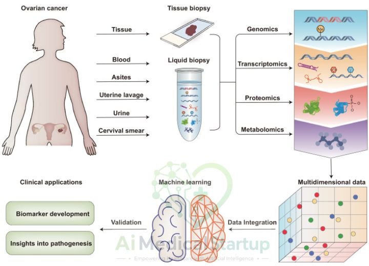
Combining advanced computational tools with biological research accelerates the discovery of targeted therapies for complex pulmonary conditions. Integrated Multi-omics (genomics, transcriptomics, proteomics) analysis links unique molecular signatures directly to real-world disease expressions. These computational systems identify personalized treatment options tailored to specific biological subgroups.
Translational research pipelines reveal hidden pathological mechanics across rare parenchymal lung conditions. Computational models streamline drug discovery by matching novel pharmaceutical compounds to target proteins quickly.
Multi-Omics and Biomarker Discovery
Genomics, Transcriptomics, and Proteomics Integration
Data processing models combine single-cell RNA sequencing data with structural patient scans. This multi-layered analysis identifies distinct inflammatory patterns within complex patient populations, facilitating precise Biomarker discovery.
Liquid Biopsies and Non-Invasive Early Detection
Machine learning platforms evaluate circulating cell-free DNA (cfDNA) and cfRNA fragmentation patterns isolated from routine blood samples using Liquid biopsy (cfDNA,cfRNA) technology. Combined with advanced imaging, blood sample analytics detect early-stage lung cancers long before physical tumors appear on standard chest radiographs.
AI-Driven Drug Discovery and Molecule Repurposing
Virtual screening platforms model molecular docking interactions to evaluate established compounds for treating progressive fibrotic tissue conditions.
Drug discovery workflow:
Identification and 3D modeling of pathological tissue proteins
In silico computational screening of chemical compound libraries
Validation of repurposed molecules for accelerated clinical testing
Using smart software for Drug repurposing / drug discovery significantly shortens developmental timelines for orphan lung diseases.
Key Challenges and Implementation Bottlenecks
Deploying artificial intelligence models into daily healthcare workflows introduces important technical, legal, and operational hurdles. Validating predictive software across diverse populations requires strict quality controls to prevent dangerous algorithmic biases. Healthcare leaders must address these system bottlenecks before committing to full-scale clinical adoption.
Establishing clear operational standards ensures artificial intelligence tools enhance patient safety without replacing sound clinical judgment. Transparent implementation strategies build long-term confidence among healthcare providers and patients alike.
Data Quality, Heterogeneity, and Algorithmic Bias
Diagnostic algorithms trained on limited or uniform datasets often show reduced accuracy when deployed in diverse real-world clinics. Variations in scanner hardware, imaging protocols, and patient demographics introduce data noise that degrades algorithm performance. Developers must curate diverse multi-center datasets to build safe, unbiased clinical models.
Interpretability, “Black Box” Models, and Physician Trust
Complex neural networks often generate diagnostic scores without displaying their underlying reasoning steps, making physicians hesitant to adopt their recommendations. Implementing Explainable AI (XAI) / SHAP frameworks displays visual heatmaps that explain automated outputs. These visual explanations help clinicians verify algorithmic predictions with confidence.
Regulatory, Medico-Legal, and Economic Barriers
FDA/EMA Approval Pathways for Software as a Medical Device (SaMD): Securing regulatory approval for medical software requires rigorous clinical validation and data safety testing.
US Regulation
The FDA utilizes Predetermined Change Control Plans to oversee self-updating machine learning software safely.
European Standards
The EU AI Act and Medical Device Regulation mandate strict data governance, human oversight, and ongoing safety tracking.
Reimbursement Models and ROI in Primary Care
Establishing clear billing pathways is essential for long-term technology adoption in primary care. In the United States, practices rely on specific CPT billing codes to cover remote patient management and digital tracking services:
99453
Initial setup and patient education for remote monitoring equipment.
99454
Monthly device supply and automated physiological data transmission services.
99457
First twenty minutes of clinical management time spent reviewing remote patient data.
Explainability and Translational Bottlenecks in Clinical Practice
Integrating diagnostic algorithms into existing electronic health record systems often uncovers severe technical hurdles. Forcing physicians to log into separate external software portals creates workflow friction that slows adoption. Software developers must build seamless background integration to ensure rapid clinical acceptance.
Future Directions and Emerging Paradigms
The next era of digital respiratory care will feature fully integrated, privacy-focused computing models. Future diagnostic engines will process real-time patient physiological metrics alongside local environmental data seamlessly. These emerging systems will transform reactive care models into proactive, precision health management platforms.
Connecting multiple diagnostic tools will establish comprehensive continuous care networks across community clinics. Intelligent platforms will deliver personalized health guidance to clinicians and patients without interrupting daily workflows.
Federated Learning (FL) for Privacy-Preserving Research
De-centralized Federated learning (FL) models train algorithms across global hospital networks without centralizing sensitive patient records.
Federated learning workflow:
Local data training & private gradient computation
Local data training & private gradient computation
Local data training & private gradient computation
Aggregation of anonymized weights without sharing raw patient records
Updated master intelligence deployed back to participating nodes
Local patient data remains protected behind hospital firewalls while the shared diagnostic model learns from global data trends.Multimodal Pulmonary Foundation Models
Next-generation foundation architectures combine genomic profiles, high-resolution CT scans, tissue analysis from Digital pathology via Whole-slide imaging (WSI), and longitudinal lung function tracking simultaneously. Synthesizing these diverse data streams enables holistic disease profiling across all stages of patient care.
Seamless Integration into Primary Care Workflows
Future digital assistants will run quietly in the background during routine medical appointments. Highlighting relevant patient history and suggesting evidence-based test orders within native electronic records empowers family doctors to deliver specialized care effortlessly.
A Real-World Clinical Case Study: Early Diagnostic Transformation
A suburban primary care clinic implemented an AI-driven spirometry and chest radio-graph screening tool to improve local respiratory triage. A 58-year-old patient presented with mild, non-specific exertional breathlessness, initially suspected to be age-related reconditioning.
The integrated software analyzed the patient’s flow-volume spirometry loop, flagging an early restrictive pattern that standard manual inspection missed. Concurrently, the automated imaging algorithm identified subtle lower-lobe reticular opacities on a routine chest X-ray, suggesting early interstitial changes.
The combined clinical alert prompted an early referral to a regional pulmonology center. High-resolution computed tomography confirmed early-stage idiopathic pulmonary fibrosis. Starting anti-fibrotic therapy early preserved the patient’s lung function, demonstrating how intelligent primary care screening prevents diagnostic delays and improves long-term outcomes.
Frequently Asked Questions
Common inquiries about AI-driven respiratory care, remote monitoring, and data privacy.
How does artificial intelligence improve early lung cancer screening in primary care settings?
Algorithms evaluate low-dose computed tomography images automatically to highlight microscopic sub-solid nodules and track volumetric growth rates over time. This automated characterization identifies suspicious lesions earlier, accelerating life-saving interventions for high-risk patients.
Can primary care physicians rely on automated spirometry readings safely?
Yes, integrated diagnostic platforms analyze flow-volume loops alongside global lung function standards instantly. They alert clinicians to patient effort artifacts and flag early obstructive patterns like emphysema with high technical accuracy.
What is the role of remote patient monitoring in managing chronic respiratory conditions?
Wearable sensors continuously track pulse oximetry, respiratory rates, and daily cough frequency from home. Care teams review these physiological trends remotely to spot impending asthma or emphysema flare-ups before emergency hospital visits occur.
How do regulators protect patient privacy when training medical algorithms?
Privacy-preserving frameworks like decentralized federated training allow models to learn across multiple institutions by sharing encrypted algorithm updates rather than raw patient charts. Additionally, stringent regulatory frameworks enforce strict data governance standards.
Conclusions
Integrating artificial intelligence into primary respiratory care marks a major shift toward proactive, personalized pulmonary medicine. Intelligent software tools reduce clinician workloads, eliminate diagnostic delays, and expand specialized screening capabilities into routine primary care consultations. Overcoming regulatory, technical, and economic challenges will establish these diagnostic tools as essential components of modern healthcare systems. The future of lung healthcare relies on collaboration between advanced digital tools and human clinical intuition. Adopting these connected technologies ensures medical teams treat chronic lung conditions earlier, more accurately, and with unprecedented precision. Primary care centers equipped with intelligent diagnostic tools will drive the next generation of improved patient outcomes worldwide.

Dr. Javeria is a Resident Pulmonologist and Chest Specialist with clinical experience across Basic Health Units and tertiary healthcare settings. Her professional focus is centered on respiratory health, pulmonary care, and chest-related conditions, bringing practical clinical insight from both primary and tertiary healthcare environments.
