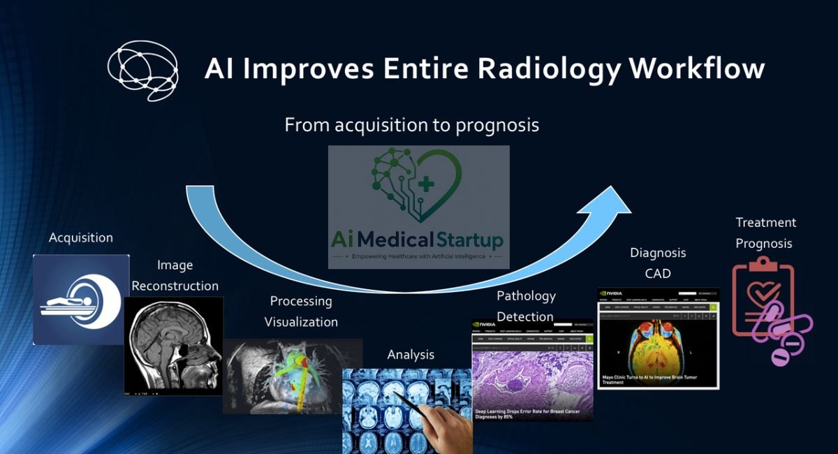
Best AI Tools in Radiology

Best AI Tools in Radiology: Top Solutions for Medical Imaging in 2026
Radiology is changing quickly as technology moves from research labs into everyday clinical practice. In 2026, AI in radiology is helping healthcare teams analyze images, prioritize urgent cases, automate measurements, and support faster clinical decisions. From X-rays and CT scans to MRI and mammography, medical imaging AI is becoming an important part of modern diagnostic workflows. But with so many platforms available, finding the best AI tools in radiology can feel overwhelming.
Each solution has different strengths, regulatory requirements, clinical evidence, and workflow capabilities. This guide explores leading radiology AI solutions, their applications, benefits, limitations, and real-world uses, helping you understand which technologies may best fit modern radiology practices and healthcare organizations.
What Are AI Tools in Radiology?
AI in radiology refers to software that uses machine learning, deep learning, computer vision, or related technologies to analyze medical imaging. These systems can identify patterns that may be difficult or time-consuming to evaluate manually. Depending on their design, they may support detection, classification, image segmentation, measurement, triage, reconstruction, or reporting.
Modern radiology AI is best understood as a collection of specialized technologies rather than one universal system. Some applications analyze a chest X-ray for suspicious findings. Others analyze a CT scan for intracranial hemorrhage or stroke. Some support MRI reconstruction, while others help create automated radiology reports. The common goal is to give clinicians useful information at the right point in the workflow.
How artificial intelligence is used in medical imaging
The basic process is surprisingly straightforward. An imaging study enters the hospital’s digital workflow, usually through systems connected to PACS, RIS, or other clinical infrastructure. An AI application processes the relevant images and searches for predefined patterns. Depending on the product, it may then highlight a finding, calculate a measurement, prioritize the case, or provide another form of clinical decision support.
This is where AI for medical image analysis becomes valuable. A system may recognize a suspected fracture, identify a possible pneumothorax, measure a Cobb angle, or flag a CT study for urgent review. The output does not automatically become the final diagnosis. Instead, the radiologist evaluates the AI result alongside the images, patient history, prior studies, and other clinical information.
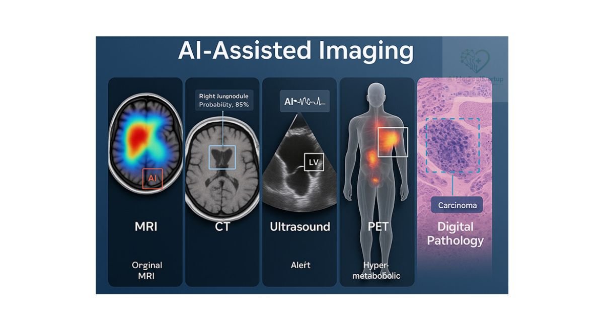
AI vs traditional radiology software
Traditional radiology software mainly helps clinicians store, retrieve, display, transmit, and document images. A PACS, for example, provides the environment in which images can be viewed. DICOM allows medical images and related information to move between compatible systems. These technologies remain fundamental to modern diagnostic imaging.
AI adds another layer. Instead of simply displaying an image, AI software for radiologists can analyze it and produce a clinically relevant output. That output might be a detection marker, probability score, measurement, segmentation, or worklist alert. In this sense, AI-powered radiology complements traditional infrastructure rather than replacing it.
Machine learning and deep learning in radiology
Machine learning in radiology allows computers to identify relationships within data and use those relationships to generate predictions or classifications. Deep learning takes this further through multilayered neural networks that can learn complex visual patterns. Many modern medical-image applications use deep-learning architectures because they can process large volumes of imaging data.
Training quality matters enormously. An AI model can perform impressively in the dataset used for development but behave differently in another hospital. Scanner manufacturers, acquisition protocols, patient demographics, disease prevalence, image quality, and clinical workflows can all affect performance. Therefore, strong clinical validation is essential before assuming that a model will perform equally well everywhere.
Generative AI and large language models in radiology
Generative AI introduces a different category of capability. Instead of only identifying visual patterns, systems using natural language processing and large language models can work with clinical text. They may help summarize patient information, draft impressions, organize findings, or support structured radiology reporting.
However, language models introduce their own risks. A fluent sentence can still be clinically wrong. Recent FDA discussions have highlighted the importance of validation and human review for generative AI used in healthcare. In one FDA-discussed evaluation, clinically significant errors in AI-generated radiology impressions fell from 4.8% before review to 1% after radiologist editing. The lesson is simple: polished language is not the same as clinical accuracy.
What AI can and cannot do for radiologists
AI can process images rapidly, identify predefined abnormalities, automate repetitive measurements, and help prioritize urgent cases. It can also provide a second layer of analysis that supports AI-assisted diagnosis. These capabilities can be particularly useful in high-volume environments where hundreds or thousands of studies may need attention.
AI cannot independently understand every clinical situation. It may struggle with unusual pathology, artifacts, incomplete information, unexpected anatomy, or cases outside its validated population. It also cannot replace the broader reasoning of a clinician who integrates imaging with symptoms, history, laboratory results, previous examinations, and treatment plans. Human oversight therefore remains central to responsible deployment.
Benefits of AI in Radiology
The benefits of AI in radiology extend beyond faster image interpretation. A well-designed system can influence what happens before, during, and after image review. It can help identify urgent examinations, automate measurements, support reporting, and reduce repetitive tasks. The greatest value often appears when AI is designed around a real clinical bottleneck rather than added simply because a hospital wants to “use AI.”
The practical advantage is workflow optimization. Imagine a busy emergency department where several critical CT studies arrive within minutes. A validated AI system may flag examinations containing potentially urgent findings and help move them toward the appropriate clinical queue. It does not remove the radiologist from the process. It helps the right human see the right case sooner.
Faster diagnosis in high-pressure settings
Time matters greatly in emergency radiology. Conditions such as stroke, intracranial hemorrhage, pulmonary embolism, and major trauma can require rapid action. AI tools for emergency radiology can analyze incoming studies and identify examinations that may need urgent attention.
The value is often measured in workflow time rather than a simple “AI versus human” accuracy contest. An AI triage system may work continuously in the background while clinicians manage other responsibilities. When an urgent case is detected, the system can generate an alert or change its priority. The final interpretation still belongs to the clinical team.
Improved diagnostic accuracy and consistency
AI may provide an additional opinion during image interpretation. This can be useful when the finding is subtle or when a department wants an additional quality-control layer. However, it would be misleading to say that AI automatically improves every diagnosis.
Performance depends on the algorithm, disease, modality, population, and implementation. A system trained to identify one abnormality may have little value for another. Diagnostic accuracy should therefore be evaluated for the specific clinical task. Hospitals should examine sensitivity, specificity, false-positive rates, external validation, and relevant clinical outcomes rather than relying on a single marketing number.
Enhanced decision support for radiologists
Clinical decision support can provide useful information without attempting to make the entire clinical decision. An AI system might highlight suspicious areas, provide a quantitative measurement, or suggest that a study deserves closer review.
This can be particularly useful for complex workflows. A radiologist can combine AI output with visual inspection and clinical context. In that model, AI becomes another source of evidence. The clinician remains responsible for deciding whether the finding is real, relevant, and clinically meaningful.
Earlier detection of abnormalities
Some AI systems are designed to detect subtle abnormalities at an early stage. This is especially relevant to screening and high-volume imaging. A computer can analyze thousands of images without becoming tired or losing concentration.
Still, early detection should not be confused with definitive diagnosis. For example, AI may identify a suspicious lung lesion or breast abnormality. Further imaging, comparison with prior studies, clinical assessment, or tissue diagnosis may still be necessary. The AI output is a signal for attention, not automatically the final answer.
Radiology workflow optimization
Radiology workflow automation can reduce friction throughout an imaging department. AI can analyze examinations immediately after acquisition, prioritize cases, perform measurements, and support reporting.
The strongest systems fit naturally into existing workflows. A radiologist should not need to open five separate applications just to see an AI result. Integration with PACS, RIS, and other hospital systems can determine whether an otherwise excellent technology becomes genuinely useful.
Reduced repetitive administrative tasks
Reporting creates a substantial amount of repetitive work. AI can assist with AI radiology reporting, report templates, impression drafting, follow-up recommendations, and clinical documentation.
The goal is not simply to generate more text. Good automated radiology reports should help the radiologist communicate findings accurately and efficiently. Generated content still requires review because language models and automated systems can produce false positives, false negatives, omissions, or incorrect statements.
Improved patient triage and prioritization
AI triage is particularly valuable when the order in which studies are reviewed matters. A system can analyze incoming images and identify studies that may contain time-sensitive abnormalities.
The FDA recognizes radiological computer-assisted prioritization as a distinct medical-device category. These systems can prioritize time-sensitive imaging for review based on image analysis. Importantly, some triage products provide prioritization rather than a complete diagnostic interpretation.
What Are the Best AI Tools in Radiology in 2026?
There is no single winner among the best AI tools in radiology. Different platforms solve different problems. Aidoc and Viz.ai, for example, are strongly associated with acute-care workflows. Gleamer has important musculoskeletal applications. Qure.ai and Lunit have significant imaging-analysis capabilities. Rad AI focuses heavily on reporting and productivity, while Subtle Medical is known for image enhancement.
The following table provides a practical starting point. Product capabilities and regulatory indications can change, so this should be treated as a high-level comparison rather than a substitute for current vendor documentation or regulatory databases.
| AI platform | Main strength | Common imaging focus | Typical workflow role |
| Aidoc | Acute-care AI and triage | CT, X-ray and other modalities | Detection and prioritization |
| Gleamer | Musculoskeletal imaging | X-ray | Fracture and orthopedic analysis |
| Qure.ai | Automated image analysis | X-ray and CT | Detection and screening |
| Lunit | Chest and breast imaging | X-ray and mammography | Detection and decision support |
| RapidAI | Stroke imaging | CT, CTA, CTP, MRI | Stroke assessment and triage |
| Viz.ai | Acute-care coordination | CT and vascular imaging | Detection, notification and workflow |
| Annalise.ai | Broad radiology analysis | Chest X-ray and CT | Multi-finding detection |
| Rad AI | Reporting productivity | Radiology workflow | Reporting and documentation |
| Subtle Medical | Image enhancement | MRI and other imaging | Reconstruction and scan optimization |
| Arterys | Quantitative imaging | MRI, CT and cardiovascular imaging | Segmentation and analysis |
Aidoc
Aidoc is one of the prominent names in enterprise radiology AI, particularly for acute-care detection and workflow. Its technology is designed to analyze imaging studies and support the identification and prioritization of potentially urgent findings. Its main advantage is breadth. Instead of focusing on one narrow imaging problem, the platform has developed a wider ecosystem of algorithms. That makes it relevant to hospitals seeking centralized AI-powered diagnostic imaging and workflow support.
Gleamer
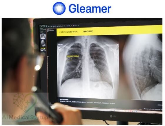
Gleamer focuses strongly on musculoskeletal and trauma imaging. Its technology is particularly relevant to AI for fracture detection, where X-ray examinations are common and rapid interpretation can be valuable. For orthopedic and emergency environments, this makes Gleamer an interesting option. The broader category of fracture detection software can help identify suspected abnormalities that deserve attention. However, clinicians should still evaluate the product’s specific indications, evidence, and regulatory status before implementation.
Qure.ai
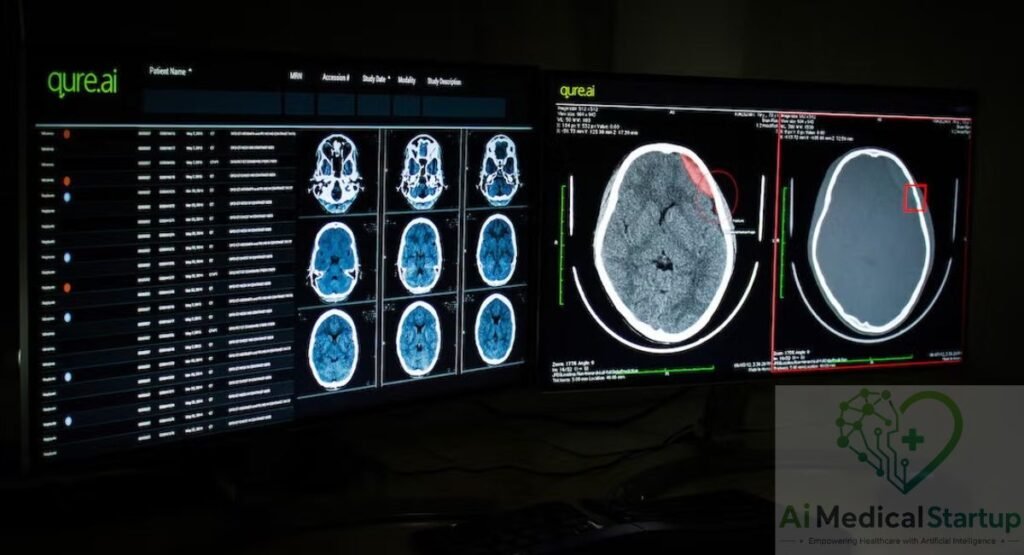
Qure.ai develops AI medical imaging solutions for several clinical applications, including chest X-ray and CT analysis. Its technology has been used for automated detection and screening workflows. One reason Qure.ai stands out is its focus on high-volume imaging environments. Automated analysis can support screening programs and help clinicians process large numbers of studies. Its regulatory record is also evolving. For example, the FDA’s 2026 AI-enabled device list includes Qure.ai’s qXR-Detect under a radiology authorization.
Lunit
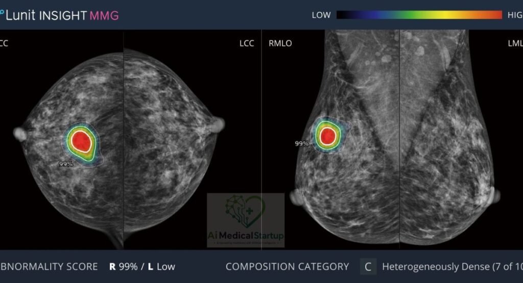
Lunit is particularly associated with chest and breast imaging. Its solutions support AI for chest X-ray analysis and breast-imaging workflows, including mammography-related applications. The platform is relevant to hospitals and screening programs that want AI-assisted detection. Lunit’s continued regulatory activity also illustrates how rapidly the medical-AI market is developing. The FDA’s current AI-enabled device list includes Lunit INSIGHT DBT, with a final decision recorded in March 2026.
RapidAI
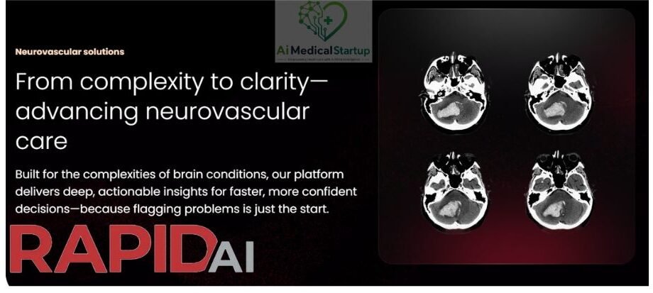
RapidAI specializes in stroke and neurovascular care. Its systems are designed around the reality that stroke treatment can depend heavily on time and accurate imaging assessment. The platform can support analysis of CT, CT angiography, CT perfusion, and related studies depending on the product. Its strongest differentiator is therefore not simply “AI image analysis.” It is the connection between imaging, patient triage, clinical communication, and time-sensitive stroke care.
Viz.ai
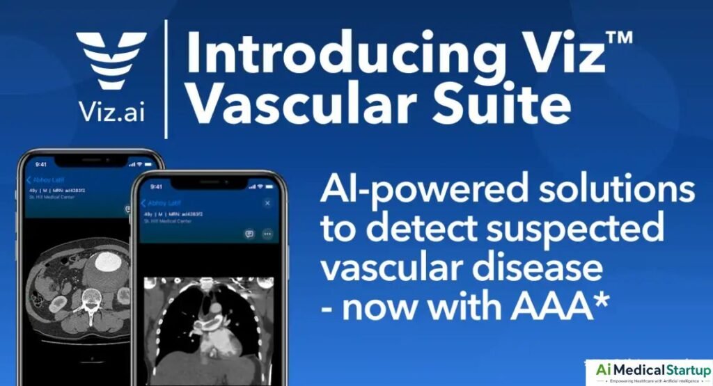
Viz.ai takes a broader acute-care approach. Its platform connects imaging analysis with notifications and clinical workflows, particularly in stroke and cardiovascular care. That distinction matters. A hospital does not necessarily need another image viewer. It may need a system that recognizes a potentially serious finding, alerts the appropriate team, and supports the movement of a patient through a time-critical pathway. Viz.ai is particularly relevant to this workflow-centered model.
Annalise.ai
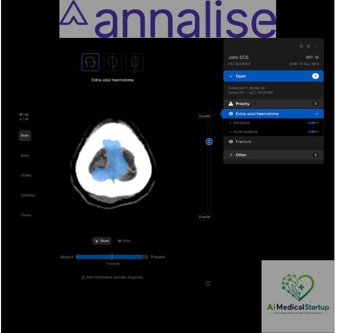
Annalise.ai develops broad AI systems for medical imaging, with notable applications in chest X-ray and head CT. Rather than focusing only on one finding, its technology can evaluate multiple potential abnormalities. This makes the platform relevant to general radiology environments. Multi-finding systems can be useful when clinicians want a broader second-reader function. However, more findings also create more opportunities for unnecessary alerts, so hospitals should carefully evaluate the balance between sensitivity and specificity.
Rad AI
Rad AI takes a different approach. Its core value is heavily connected to AI radiology reporting and radiologist productivity rather than only image detection.
Reporting tools can assist with impressions, follow-up recommendations, and repetitive documentation. This category is becoming increasingly important because radiology workloads involve far more than looking at pictures. The strongest systems reduce documentation friction while keeping the radiologist firmly in control of the final report.
Subtle Medical
Subtle Medical focuses on AI-powered image enhancement and reconstruction. This places it in a different category from fracture or stroke detection platforms.
For MRI and other modalities, image-enhancement AI can potentially improve efficiency, image quality, or acquisition workflows. The benefit may therefore appear earlier in the imaging pathway rather than at the moment of diagnosis. It is a useful reminder that medical imaging AI is much broader than disease detection.
Arterys
Arterys has focused on cloud-based medical imaging analysis and quantitative applications, including cardiovascular imaging. Its technology illustrates another important category of AI for medical image analysis: automated segmentation and measurement.
Quantitative imaging can be valuable when clinicians need reproducible measurements rather than only a simple “abnormal” or “normal” label. This can support research, treatment planning, and longitudinal patient monitoring.
Top Use Cases of AI in Radiology
The applications of AI in radiology cover almost every stage of modern imaging. Some systems analyze images for abnormalities. Others perform measurements. Some improve image quality. Others organize worklists or assist with reports.
The most useful application depends on the clinical problem. A stroke center may prioritize neurovascular triage. An orthopedic emergency department may care more about AI for fracture detection. A breast-screening service may need mammography AI. A busy outpatient imaging group may gain more value from reporting automation.
Detecting and classifying brain tumors
AI can support AI brain tumor detection, segmentation, classification, and quantitative assessment. MRI is particularly important because it provides detailed soft-tissue information.
Research applications can include brain tumor classification, tumor volume measurement, and characterization of lesions. Systems may also support research into glioma, meningioma, and other brain tumors. However, classification should not be interpreted as a substitute for pathology when tissue diagnosis is required.
Stroke and intracranial hemorrhage detection
Stroke is one of the strongest use cases for AI because treatment decisions can be highly time-sensitive. AI can analyze CT and vascular imaging for findings associated with stroke or hemorrhage.
A well-designed system can support AI triage by flagging potentially urgent examinations. The clinical value comes from shortening the path between image acquisition, recognition, communication, and treatment. The technology is therefore closely tied to workflow rather than image interpretation alone.
Fracture and trauma detection
Fracture detection is a natural application for computer vision. X-ray examinations are common, and some fractures can be subtle.
AI can assist with suspected fracture, dislocation, and other trauma findings. The output can act as an additional check for the clinician. It should not be treated as proof that a fracture is present or absent.
Chest disease and lung abnormality detection
Chest imaging offers a broad field for AI. Algorithms can assist with findings such as pneumothorax, pleural effusion, pulmonary edema, consolidation, and other abnormalities.
Chest X-ray AI is especially useful in high-volume settings. It can support screening, triage, and second-reader workflows. Still, the meaning of an abnormality depends on the patient’s symptoms and clinical context, so automated detection must be interpreted carefully.
Cancer screening and lesion detection
AI is increasingly used in cancer-related imaging. Mammography is one major area, while lung CT and other modalities offer additional opportunities.
An AI system may identify a suspicious lesion, estimate a probability, or highlight an area for review. That does not mean it has diagnosed cancer. Definitive diagnosis may require additional imaging, biopsy, pathology, or other clinical evaluation.
Radiation dose optimization
AI can contribute to radiation dose optimization by supporting image reconstruction, acquisition decisions, and image-quality management.
The goal is not simply to reduce dose at any cost. The image must remain diagnostically useful. A very low-dose scan that cannot answer the clinical question is not necessarily a successful optimization. AI can help find a better balance between radiation exposure and image quality.
Automated image segmentation and measurements
Image segmentation allows software to identify specific anatomical structures or regions within an image. Once a structure is segmented, the system can calculate volume, area, length, angle, or other measurements.
This is valuable for orthopedic AI, oncology, cardiovascular imaging, and treatment planning. Automated measurements can also improve diagnostic consistency by reducing variation caused by manual calculations.
Radiology reporting and documentation
Reporting is becoming one of the most visible uses of generative AI. Systems can help organize findings, draft impressions, create summaries, or suggest follow-up language.
The opportunity is significant, but so is the risk. A generated report may sound professional while containing a subtle error. Therefore, automated radiology reports should remain subject to careful review, editing, and clinical accountability.
Patient prioritization and triage
AI can analyze studies and identify those that may require faster review. This is especially useful in emergency and acute-care settings.
The important distinction is between diagnosis and prioritization. Some AI systems are specifically designed to move a potentially urgent study higher in a queue. FDA classifications recognize radiological computer-assisted triage and notification as a specific type of software device.
AI Applications in Radiology by Imaging Modality
AI behaves differently across imaging modalities. X-ray produces relatively standardized two-dimensional images. CT creates volumetric datasets. MRI provides rich soft-tissue information and can involve lengthy acquisition protocols. Ultrasound depends heavily on operator technique.
For that reason, the best AI tools for medical imaging should always be evaluated within their modality. A highly effective X-ray algorithm does not automatically make an excellent MRI solution. The data, clinical problem, workflow, and validation requirements are different.
| Imaging modality | Common AI applications | Main opportunity |
| X-ray | Fracture and chest analysis | High-volume detection |
| CT | Stroke, trauma, nodules, organ analysis | Rapid 3D assessment |
| MRI | Reconstruction, segmentation, tumor analysis | Quality and efficiency |
| Ultrasound | Measurements and image guidance | Operator support |
| Mammography | Lesion detection and screening | Breast cancer support |
| PET/nuclear medicine | Segmentation and quantification | Oncology and precision imaging |
AI for X-ray diagnostics
X-ray remains one of the most practical areas for AI for X-ray applications. The modality is widely available and produces large volumes of examinations.
AI can support fracture detection, chest abnormality detection, measurements, bone-age estimation, and quality assessment. The technology is especially attractive in emergency departments, outpatient imaging centers, and screening environments.
AI for CT scans
AI for CT scans covers a wide range of applications. These include stroke and hemorrhage detection, pulmonary embolism analysis, lung-nodule detection, trauma assessment, organ segmentation, and quantitative measurements.
CT also produces three-dimensional information. This gives AI more data to analyze but creates greater computational complexity. A successful CT system must therefore handle large datasets efficiently while maintaining reliable performance.
AI for MRI
For MRI, AI includes both diagnostic and image-acquisition applications. It can assist with segmentation, reconstruction, image enhancement, quantitative analysis, and lesion characterization.
One major opportunity is scan acceleration. If AI can help reconstruct useful images from fewer or faster acquisitions, patients may spend less time in the scanner. The clinical value depends on maintaining sufficient image quality for the intended diagnostic task.
AI for ultrasound
Ultrasound creates unique AI challenges because image quality can depend heavily on the operator. Probe position, angle, pressure, patient anatomy, and acquisition technique can all influence the image.
AI can support automated measurements, image-quality checks, cardiac assessment, and workflow guidance. In this environment, AI may become particularly useful as an assistant to standardize parts of the examination.
AI for mammography
Mammography is one of the most important areas for AI-supported cancer screening. Algorithms can analyze images for suspicious masses, calcification, asymmetries, and other findings.
The role of AI varies between products. Some systems provide detection support. Others may support risk assessment or workflow prioritization. Because screening involves large populations, validation across different demographic groups and imaging environments is particularly important.
AI in nuclear medicine and PET imaging
AI can assist with segmentation, image reconstruction, lesion analysis, and quantitative assessment in nuclear medicine and PET.
Oncology is an important area because PET imaging can provide information about metabolic activity. AI may help identify lesions and calculate quantitative characteristics. These technologies could eventually support more personalized assessment of treatment response.
AI for X-Ray Diagnostics: What Can It Detect?
X-ray is often the first modality people associate with radiology AI. That makes sense. X-ray examinations are fast, inexpensive compared with many advanced modalities, and widely used. They also generate enormous amounts of data.
However, “AI can read X-rays” is too broad a statement. Different systems are trained for different findings. One may focus on fractures. Another may analyze the chest. A third may perform orthopedic measurements. The safest approach is always to ask what the algorithm was specifically designed and validated to detect.
Fracture and trauma detection
AI can analyze musculoskeletal X-rays for suspected fractures and other trauma-related findings. Some systems can also flag possible dislocation or related abnormalities.
The value is strongest when the technology fits the clinical workflow. In a busy emergency department, an AI flag can encourage rapid review of a suspicious image. It does not eliminate the need for a complete clinical and radiological assessment.
Chest pathology detection
Chest AI can evaluate X-rays for several common abnormalities. Depending on the system, this may include pneumothorax, pleural effusion, consolidation, edema, or other findings.
A chest abnormality can have multiple possible causes. Therefore, AI detection is only one part of the diagnostic process. The radiologist must determine what the finding means in the patient’s clinical context.
Osteo-articular measurements
AI can automate measurements that traditionally require manual work. These may include Cobb angle, alignment measurements, limb-length calculations, and other orthopedic assessments.
Applications can include scoliosis, hallux valgus, leg length discrepancy, hip dysplasia, and femoroacetabular impingement assessment. Automated measurement can improve speed and reproducibility, especially when the same measurements are repeated over time.
Pediatric bone age assessment
Pediatric bone age assessment is another established application area. AI can evaluate hand and wrist radiographs to estimate skeletal maturity.
Traditional assessment can involve comparison with references such as the Greulich and Pyle atlas. AI can automate parts of this process by analyzing developmental features and ossification centers. The result remains an estimate rather than an absolute biological age.
Lung and cardiac abnormality detection
Chest AI may also assist with lung and cardiac findings. Examples include lung nodules, pulmonary edema, and cardiomegaly.
These systems can help prioritize or support image review. They should not be treated as universal disease detectors. Performance depends on the exact finding, patient population, image quality, and algorithm.
Automated image quality assessment
Poor-quality images can lead to diagnostic difficulty. AI can help identify positioning problems, motion, exposure issues, or other acquisition concerns.
This creates an opportunity before diagnosis even begins. If a system detects that an image is inadequate, the imaging team may be able to repeat the study when clinically appropriate. This can improve workflow and reduce avoidable delays.
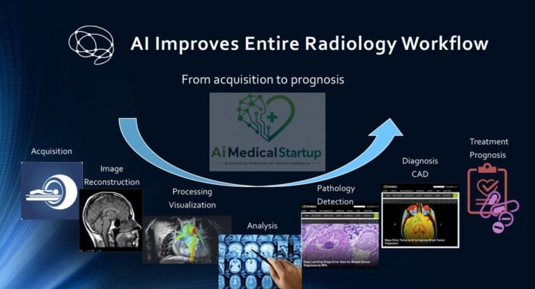
How AI Improves the Radiology Workflow
A radiology department is more than an image-reading room. It is a connected system involving scanners, technologists, scheduling, PACS, RIS, reporting, clinicians, and patients. AI becomes valuable when it improves this entire chain.
Workflow optimization can happen at several points. AI can analyze an image immediately, prioritize urgent studies, calculate measurements, assist with reports, and communicate results. The best implementation feels almost invisible because it works inside the tools clinicians already use.
Automated image analysis
Automated image analysis allows software to process images immediately after acquisition. Depending on the algorithm, it can identify suspicious patterns, segment anatomy, or calculate quantitative features.
This can create a useful second layer of review. The system does not need to replace the radiologist. Instead, it can perform repetitive computational work while the clinician focuses on interpretation and decision-making.
Worklist prioritization and triage
A normal worklist may treat examinations largely according to operational rules. AI can add another dimension by identifying potentially urgent imaging findings.
This AI triage can be valuable when a serious abnormality is hidden within a large queue. The system can help move that examination toward faster review. This is especially relevant in emergency and stroke workflows.
Clinical decision support
AI can provide additional information during clinical interpretation. It may highlight a region of interest, produce a measurement, or show an algorithmic probability.
This is clinical decision support, not necessarily autonomous diagnosis. The clinician must decide how much weight to give the AI result. A good interface should make it easy to compare AI output with the underlying image.
Automated measurements
Measurements are a perfect example of a repetitive task that computers can handle efficiently. AI can identify anatomical landmarks and calculate distances, areas, volumes, and angles.
In orthopedic imaging, this may include the Cobb angle or limb-length measurements. In cardiovascular imaging, it may involve chamber volumes or functional parameters. Automation can save time while improving consistency.
Structured reporting assistance
Structured radiology reporting helps standardize how findings are communicated. AI can assist by suggesting terminology, organizing findings, or drafting sections of a report.
The strongest approach keeps the clinician in charge. AI should make reporting easier without turning the report into an unchecked machine-generated document. This is especially important when using generative AI and NLP.
Integration with PACS and RIS
Integration can make or break an AI deployment. If clinicians have to leave their normal workflow every time they want to view an AI result, adoption may be poor.
Good integration can use PACS, RIS, HIS, DICOM, and relevant interfaces to move information between systems. Hospitals should evaluate latency, reliability, authentication, auditability, and how AI findings appear within the existing reading workflow.
Reducing radiologist administrative workload
Administrative work can consume valuable time. AI can help with documentation, report drafting, follow-up reminders, and communication. This does not mean every administrative task should be automated. The goal is to remove repetitive work while preserving accuracy and clinical accountability. A small reduction in documentation burden across thousands of examinations can become a meaningful operational benefit.
How Accurate Are AI Tools in Radiology?
Accuracy is one of the most misunderstood parts of medical AI. A vendor may advertise an impressive percentage, but that number alone tells you very little. You need to know what disease was tested, which images were used, how many patients were included, what the reference standard was, and whether the system was tested outside its development dataset.
A clinically useful evaluation should examine sensitivity, specificity, positive and negative predictive values, AUC, false-positive rates, and workflow outcomes. It should also consider whether the algorithm improves performance when used by clinicians. An AI system with excellent standalone performance may provide little practical benefit if it creates excessive alerts or disrupts workflow.
AI sensitivity and specificity
Sensitivity measures how effectively a system identifies cases that truly contain the target condition. High sensitivity can be important when missing a serious abnormality has significant consequences.
Specificity measures how well the system avoids incorrectly labeling normal cases as positive. The balance matters because increasing sensitivity can sometimes increase false positives. Hospitals should therefore evaluate both measures rather than choosing a product based on one impressive statistic.
Comparing AI performance with radiologists
Comparisons between AI and clinicians require careful interpretation. A computer may perform differently from a human when given only images, while a radiologist can consider clinical history and previous examinations.
The more meaningful question is often whether AI-assisted diagnosis improves human performance. If radiologists work more accurately or efficiently with the tool than without it, that may be more clinically useful than proving that an algorithm can outperform a clinician under artificial test conditions.
Clinical validation and real-world evidence
Clinical validation should ideally extend beyond internal testing. External datasets, prospective studies, independent evaluations, and real-world deployments can reveal problems that development datasets miss.
Peer-reviewed research is particularly useful, but publication alone does not guarantee clinical usefulness. You should examine study design, patient population, sample size, comparator, reference standard, and whether the reported results match the intended use of the product.
False positives and false negatives
Every AI system can make errors. A false positive occurs when the system flags something that is not actually present. A false negative occurs when it fails to identify a condition that is present.
The clinical consequences differ by use case. A false positive in a low-risk screening workflow may create additional work. A false negative in an emergency setting may delay treatment. Product evaluation should therefore consider the clinical consequences of each type of error.
Why AI performance varies between hospitals
AI performance can change when the environment changes. Different hospitals may use different scanners, acquisition protocols, patient populations, contrast protocols, or image-processing pipelines.
This is sometimes described as dataset shift or distribution shift. An algorithm that performs well in one environment may need additional validation elsewhere. That is why local testing and continuous monitoring can be important even after regulatory authorization.
Understanding AUC, sensitivity, specificity and other metrics
AUC describes how well a model separates positive and negative cases across different classification thresholds. Sensitivity and specificity describe performance at a selected threshold. Positive predictive value depends heavily on disease prevalence, while negative predictive value also changes with prevalence.
For procurement teams, the best metric depends on the task. A triage system may prioritize sensitivity for serious conditions. A measurement system may require very low measurement error. A reporting system may need different quality measures altogether. There is no single number that defines AI quality.
FDA-Cleared and Regulated AI Tools for Radiology
Regulation is essential because medical AI can influence diagnosis, treatment, and patient care. In the United States, the FDA maintains an AI-enabled medical-device list designed to identify AI-enabled devices authorized for marketing. The agency says listed devices have met applicable premarket requirements, including review of safety and effectiveness appropriate to their intended use.
The regulatory landscape is also changing. The FDA has continued developing guidance around AI-enabled devices, including lifecycle management and predetermined change-control approaches. In August 2026, the agency also opened a discussion on regulatory considerations for generative-AI-enabled medical devices. For hospitals, this means regulatory status should be checked regularly rather than treated as a permanent label.
What does FDA clearance mean?
FDA clearance generally means the agency has determined that a device meets the applicable requirements for its pathway and intended use. For many radiology AI products, the relevant pathway is 510(k).
The important point is that clearance applies to a specific medical device and its intended use. It does not mean the software can safely perform every possible imaging task. The FDA’s current classification system includes radiological AI software categories covering automated image processing and analysis.
FDA-cleared vs FDA-approved AI software
“Cleared” and “approved” are not interchangeable terms. FDA clearance commonly refers to the 510(k) pathway, while approval is associated with a different regulatory pathway and evidence standard.
When evaluating FDA-cleared AI tools for radiology, check the exact device name, submission number, intended use, indication, and authorization pathway. Do not assume that a company-wide statement such as “FDA-cleared technology” applies to every algorithm sold by that company.
CE marking and European regulation
The European market has its own regulatory requirements. CE marking indicates conformity with applicable European requirements, but it should not be treated as a simple equivalent of an FDA label.
The EU AI Act also introduces a risk-based framework. Certain AI systems connected to medical devices can fall into high-risk categories and face requirements involving risk management, data quality, information, and human oversight. European hospitals should therefore evaluate both medical-device conformity and relevant AI governance obligations.
UK and other international regulatory considerations
In the UK, software and AI used for medical purposes may be regulated as medical devices. The MHRA states that many software and AI products used in health and social care fall within medical-device regulation.
The UK framework continues to evolve. In September 2026, the government tabled proposed amendments related to medical-device regulation and the future development of a medical-device licensing regime. Hospitals should therefore verify current requirements rather than relying on outdated assumptions about UKCA, CE, or transitional arrangements.
Why regulatory status matters when choosing an AI tool
Regulatory status helps establish whether a product is authorized for a particular medical purpose in a particular market. It does not answer every procurement question, but it is an important starting point.
A hospital should also examine evidence, cybersecurity, interoperability, intended use, user training, post-market monitoring, and clinical governance. Regulatory authorization and clinical suitability are related, but they are not identical concepts.
Challenges, Limitations and Ethical Issues of AI in Radiology
The challenges of AI in radiology are not purely technical. Some involve clinical judgment, patient rights, workflow design, and responsibility. A highly accurate algorithm can still create problems if it generates too many alerts, does not integrate with PACS, or is used outside its intended population.
Trust is another major issue. Clinicians need to understand what an AI system does, where it performs well, and where it can fail. Patients also need confidence that their images and health information are handled appropriately. Responsible AI-powered radiology therefore requires governance alongside technology.
False-positive and false-negative results
No AI system is perfect. False-positive results can increase workload and unnecessary follow-up. False-negative results can create a false sense of reassurance.
The correct threshold depends on the application. Emergency triage may tolerate more false positives if it helps minimize missed critical findings. Other applications may require greater specificity. The important point is to evaluate error rates in relation to actual clinical consequences.
Algorithmic bias and health disparities
Algorithmic bias can occur when training data do not adequately represent the population in which the system is deployed. Differences in age, sex, ethnicity, disease prevalence, scanner technology, or healthcare access can affect performance.
This matters because a system that works well for one population may not perform equally well for another. Developers and healthcare organizations should therefore examine subgroup performance and seek evidence across diverse populations.
Patient privacy and medical data security
Medical images contain sensitive information. Hospitals must consider data privacy, access controls, encryption, authentication, storage, transmission, and vendor practices.
In the United States, HIPAA may apply to protected health information. In the EU and UK, GDPR and related data-protection requirements can be important. Healthcare data security should be evaluated before deployment, particularly when AI systems use cloud infrastructure.
AI explainability and clinical trust
Some deep-learning systems can behave like black boxes. They may produce an output without providing an explanation that makes intuitive sense to a clinician.
Explainable AI can help by providing heatmaps, highlighted regions, confidence information, or other interpretable signals. These features are useful, but visualization alone does not prove that the algorithm is correct. Explainability should support, not replace, rigorous validation.
Liability when AI makes an error
Who is responsible when AI contributes to a wrong clinical decision? The answer can involve clinicians, healthcare organizations, software vendors, and applicable legal frameworks.
This is why clinical accountability must remain clear. Hospitals should define who reviews AI results, how errors are reported, how software updates are managed, and what happens when the system is unavailable. A vague responsibility model can create safety problems.
Overreliance on automated recommendations
Humans can develop automation bias. If software repeatedly provides useful recommendations, users may gradually trust it too much.
Radiologists should therefore remain alert to contradictory findings. An AI output should be considered evidence, not unquestionable truth. Strong clinical workflows make it easy for users to inspect the original images and challenge the algorithm.
Interoperability and workflow problems
Even excellent AI can fail operationally. Slow processing, poor interfaces, incompatible systems, duplicate alerts, and difficult authentication can frustrate users.
Integration with PACS, RIS, HIS, and other infrastructure should be tested before large-scale deployment. A hospital should evaluate the complete workflow rather than judging the algorithm in isolation.
Cost and implementation challenges
The cost of AI includes more than the software license. Hospitals may need integration work, cloud or server infrastructure, cybersecurity reviews, training, support, monitoring, and maintenance.
The right question is therefore not simply “How much does this AI tool cost?” It is “What value does it produce relative to its total cost?” A product that saves significant reporting time or reduces critical delays may justify a higher price than a cheaper tool with little practical impact.
How to Choose the Best AI Tool for Radiology
Choosing the best AI software for radiologists begins with the clinical problem. Do not start by asking which vendor has the most algorithms. Start by identifying what your department wants to improve.
A procurement team should then evaluate clinical evidence, regulatory status, workflow integration, security, usability, and total cost. A tool that performs well in a research paper but creates operational problems may deliver little real-world value. The best product is usually the one that solves a clearly defined problem and fits naturally into the existing system.
| Evaluation area | What to investigate |
| Clinical use case | What exact problem does the AI solve? |
| Regulatory status | Is the product authorized for the intended use and market? |
| Evidence | Are there independent and peer-reviewed studies? |
| Accuracy | What are sensitivity, specificity, AUC and error rates? |
| Integration | Does it work with PACS, RIS and DICOM workflows? |
| Security | How are images and patient data protected? |
| Cost | What is the total cost of ownership? |
| Support | What training, monitoring and technical support are provided? |
Define your clinical use case
Start with a specific problem. You might want to reduce fracture misses, prioritize stroke studies, accelerate MRI, improve mammography screening, or reduce reporting time.
A clear use case makes evaluation much easier. It also prevents hospitals from buying broad AI platforms without a measurable clinical objective.
Check regulatory clearance
Confirm the regulatory status of the exact product. Do not rely only on a vendor’s general statement.
In the United States, review the FDA database and intended use. In Europe, examine the applicable medical-device conformity requirements and EU obligations. Review the current MHRA framework for the United Kingdom. Regulatory status can change as products and regulations evolve.
Evaluate clinical evidence
Strong clinical evidence should be central to procurement. Look for external validation, prospective evaluations, peer-reviewed research, and real-world studies.
Also examine who funded the study. Vendor-sponsored research can be useful, but independent evidence provides another perspective. A large marketing claim should never substitute for careful clinical evaluation.
Assess accuracy and performance
Look beyond overall accuracy. Evaluate sensitivity, specificity, AUC, positive predictive value, negative predictive value, and false-positive rates.
For workflow tools, also measure time saved, alert response, report turnaround, and user acceptance. Clinical AI should be judged according to the outcome it is designed to improve.
Check PACS and RIS compatibility
A good algorithm should fit the existing infrastructure. Ask whether the system supports PACS, RIS, DICOM, and other interfaces used by the hospital.
Also test the user experience. AI findings should be visible at the right moment without requiring unnecessary clicks. Integration is not a technical afterthought. It directly affects adoption.
Evaluate cybersecurity and data privacy
Ask where data are stored, how they are transmitted, who can access them, how long they are retained, and what happens if the service becomes unavailable.
Organizations should also consider applicable HIPAA, GDPR, and local cybersecurity requirements. A clinically useful AI system still needs strong patient safety and information-security controls.
Consider scalability and cost
A pilot may involve a few hundred examinations. An enterprise deployment may involve millions. The system must remain reliable as usage grows.
Cost models can vary. Vendors may charge per study, per algorithm, by subscription, or through enterprise agreements. Procurement teams should calculate integration, support, infrastructure, and maintenance costs as part of the total investment.
Review training and technical support
Users need to understand what the AI does and what it does not do. Training should explain the interface, common errors, limitations, and escalation procedures.
Technical support is equally important. Hospitals should ask how quickly problems are handled, how software updates are validated, and how performance is monitored after deployment.
The Future of AI in Radiology
The future of AI in radiology will probably involve many different forms of intelligence working together. Image-analysis systems will continue to improve, while generative AI will increasingly interact with reports, clinical records, and workflows.
The biggest shift may be from isolated algorithms to integrated clinical systems. Instead of one tool detecting one finding, future platforms may connect imaging, clinical history, laboratory data, prior examinations, and reporting. This could make AI more useful, but it will also increase the importance of validation, transparency, privacy, and human control.
Multimodal AI and medical imaging
Multimodal AI combines different types of information. Instead of analyzing only an image, a system could consider the image alongside clinical history, laboratory results, previous scans, pathology, or other data.
This approach could provide richer clinical decision-making support. However, more information also creates more opportunities for errors and hidden correlations. Multimodal systems will require strong evidence before they become routine clinical tools.
Generative AI for radiology reports
Generative AI is likely to become increasingly important in reporting. Systems can help draft impressions, summarize findings, organize information, and create patient-friendly explanations.
The major challenge is reliability. AI-generated language can sound authoritative even when it is wrong. Human review therefore remains essential. The FDA has specifically been exploring regulatory considerations for generative-AI-enabled medical devices in 2026.
AI-powered personalized imaging
Future systems may help tailor imaging protocols to individual patients. AI could potentially consider body habitus, previous examinations, clinical questions, and other factors when optimizing acquisition.
This could contribute to more personalized imaging. The goal would be to obtain the information needed for a particular patient while avoiding unnecessary acquisition time, contrast, or radiation exposure where applicable.
Predictive analytics in radiology
Radiology AI may gradually move beyond identifying what is already visible. Predictive systems could estimate future disease risk, treatment response, or patient outcomes.
This is a different task from image interpretation. Predictive analytics must be carefully validated because a statistical association does not necessarily mean that a prediction is clinically useful or causally meaningful.
Autonomous and semi-autonomous image interpretation
Some areas of imaging may eventually support greater automation. The likelihood depends on the clinical task, risk level, evidence, regulation, and consequences of error.
Low-risk repetitive measurements may be easier to automate than complex diagnostic interpretation. The future is therefore more likely to contain a mixture of autonomous and supervised systems rather than one sudden transition to fully automated radiology.
Human-AI collaboration in radiology
The strongest long-term model may be collaboration. AI can handle repetitive computational work while clinicians provide judgment, context, communication, and accountability.
That means the question may shift from “Can AI replace the radiologist?” to “How can a radiologist use AI safely and effectively?” The answer will depend on better interfaces, stronger evidence, responsible regulation, and continuous monitoring.
AI in Radiology FAQs
The rapid growth of AI in radiology has created many practical questions. Clinicians want to know which platforms are useful, patients want to know whether AI is safe, and healthcare organizations want to know whether the technology is worth the investment.
The most accurate answers depend on the clinical application. There is no universal “best” AI product, and there is no single accuracy number that applies to every algorithm. The questions below provide a practical overview.
What is the best AI tool for radiology?
There is no single best tool for every radiologist. The right choice depends on the clinical problem, imaging modality, workflow, regulatory status, evidence, and budget.
For example, a stroke center may prioritize RapidAI or Viz.ai, while a department focused on musculoskeletal X-ray may consider Gleamer. A reporting-focused organization may look more closely at Rad AI. The best solution is the one that solves your specific problem effectively and safely.
What is the most commonly used AI in radiology?
There is no single AI system that dominates every radiology workflow. Adoption is divided across detection, triage, reporting, image enhancement, measurement, and screening.
Enterprise platforms such as Aidoc and other specialized solutions are used for different clinical purposes. Market share also varies by country, hospital system, specialty, and regulatory environment.
Can AI replace radiologists?
Current clinical AI is better understood as augmentation than replacement. Many systems perform narrowly defined tasks such as detection, prioritization, segmentation, measurement, or reporting assistance.
Radiologists still provide broader clinical reasoning. They interpret findings in context, communicate with clinicians, compare prior examinations, recommend additional imaging, and take responsibility for the final interpretation. Human oversight remains an important part of safe AI deployment.
How accurate is AI in radiology?
Accuracy varies significantly. It depends on the algorithm, target condition, imaging modality, patient population, dataset, and clinical environment.
You should examine sensitivity, specificity, AUC, predictive values, false-positive rates, and external clinical validation. A vendor’s headline accuracy number should never be considered sufficient evidence by itself.
What AI tools are FDA-cleared for radiology?
The FDA maintains an AI-enabled medical-device list containing devices authorized for marketing in the United States. The list is updated periodically and includes numerous radiology products.
The exact authorization matters. For example, the FDA database contains radiology AI products under automated image-processing and triage classifications. A healthcare organization should verify the specific device, intended use, and current authorization rather than relying on a general company claim.
How much do radiology AI tools cost?
Pricing varies widely. Enterprise platforms often use customized contracts rather than simple public pricing.
The final cost can include licensing, per-study fees, integration, cloud infrastructure, training, support, monitoring, and maintenance. For that reason, procurement teams should calculate total cost of ownership instead of comparing license prices alone.
Can AI detect cancer on an X-ray?
Some AI systems can identify findings that may be associated with cancer or other suspicious abnormalities on imaging. For example, algorithms may flag a suspicious lung finding on a chest X-ray.
However, detecting a suspicious abnormality is not the same as diagnosing cancer. Definitive diagnosis may require additional imaging, clinical assessment, or pathology. AI should therefore be considered a support tool rather than a standalone cancer diagnosis system.
How is AI used in CT and MRI?
AI for CT scans can support stroke detection, hemorrhage detection, pulmonary analysis, trauma assessment, lung-nodule detection, segmentation, and quantitative analysis.
AI for MRI can support reconstruction, scan acceleration, segmentation, image enhancement, tumor analysis, and quantitative imaging. The specific capabilities depend on the individual medical device and its intended use.
Is AI safe to use in radiology?
AI can be used safely when it has appropriate evidence, regulatory authorization where required, suitable clinical governance, cybersecurity controls, and proper human oversight.
Safety also depends on how the technology is implemented. Poor workflow integration, excessive alerts, unclear responsibility, or overreliance on automated results can introduce risks. MHRA guidance similarly emphasizes safety, evidence, lifecycle management, transparency, and post-market considerations for software and AI as medical devices.
What are the disadvantages of AI in radiology?
The disadvantages include false positives, false negatives, algorithmic bias, privacy risks, cybersecurity concerns, implementation costs, interoperability problems, and automation bias.
AI also requires continuous evaluation. Models can behave differently as patient populations, scanners, protocols, and workflows change. Responsible deployment therefore requires monitoring rather than treating installation as the end of the process.
Final Takeaway: Which Are the Best AI Tools in Radiology in 2026?
The best AI tools in radiology are not necessarily the platforms with the longest feature lists. They are the systems that solve a real clinical problem, fit naturally into the existing workflow, demonstrate strong clinical evidence, and have appropriate regulatory status for the market in which they will be used.
Aidoc, Gleamer, Qure.ai, Lunit, RapidAI, Viz.ai, Annalise.ai, Rad AI, Subtle Medical, and Arterys each represent different approaches to AI-powered diagnostic imaging. Some focus on detection. Others specialize in triage, reporting, image enhancement, or quantitative analysis. Comparing them by category is therefore more useful than declaring one universal winner.
For healthcare organizations in the USA, UK, and EU, the smartest approach is to evaluate AI as a clinical technology rather than a simple software purchase. Verify the intended use. Review the evidence. Check regulatory requirements. Test the workflow. Protect patient data. Monitor performance. Most importantly, keep qualified clinicians involved in the decision-making process. The real promise of artificial intelligence in radiology is not that machines will simply replace human expertise. It is that carefully designed systems can help radiologists work faster, analyze complex information, reduce repetitive tasks, and focus more attention on the patients who need it most.

Dr. Kanza Sarfraz, M.B.B.S., is a medical doctor and graduate of Allama Iqbal Medical College, Lahore. She brings nearly seven years of clinical experience across tertiary-care hospitals, medical headquarters, and healthcare facilities in both the public and private sectors. Her clinical experience provides a practical perspective on healthcare delivery, emerging medical technologies, and the evolving role of artificial intelligence in medicine.